mail_outline sales@mediastorehouse.com
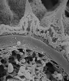
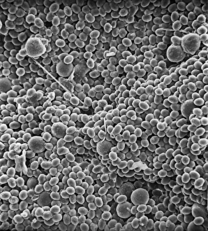
CoccolithsScanning electron microscope (SEM) image of coccoliths, these are the limestone scales surrounding the marine phytoplankton coccolithophores
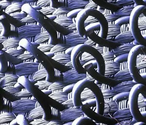
VelcroA trademarked name for a fastening tape made up of a strip of nylon with a surface of minute hooks, that fasten to another strip with a surface of uncut pile. A SEM image
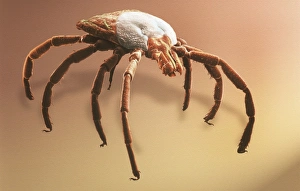
Amblyomma sp. hard backed tickScanning electron microscope view of a hard backed tick from the family Ixodidae. Coloured artificially on computer
Copper in unspecified mineralScanning electron microscope image of an elemental map showing the distribution of copper (Cu) in mineral samples
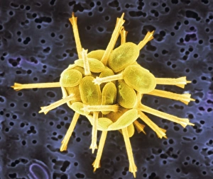
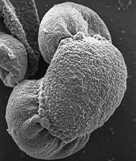
Fractured pollen grainScanning electron microscope (SEM) image showing a fractured pollen grain
Bellis perenis, daisy petalScanning electron microscope (SEM) image of a daisy petal. Published in Close-Up (2004) by Chris Jones and Alex Ball (inside cover)
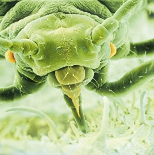
Aphis fabae, black bean aphidScanning electron microscope image showing a frontal view of a black bean aphid on leaf (x100). Aphids or plant lice are small, plant-sucking insects
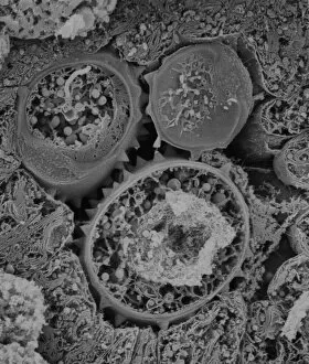
Florisphaera profundaA coccolithophore with highly modified, plate-like coccoliths. This is a very common deep dwelleing species, typically living at about 100-150m depth in the water column
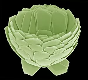
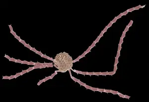
Ophiaster formosusA coccolithophore with long appendages formed of strings of highly modified coccoliths. Collected from the West Pacific. Specimen diameter 50m. False-coloured SEM image
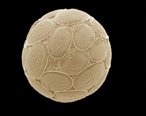
Pontosphaera japonica. A coccolithophore with relatively large, flat, coccoliths. Collected from off Hawaii. Specimen diameter 22m. False-coloured SEM image
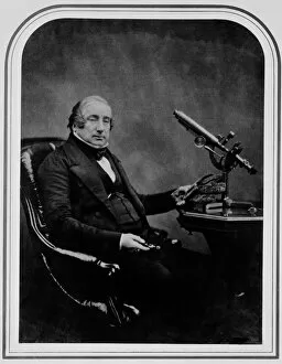
Dr James Scott Bowerbank (1797-1877)Portrait of Dr James Scott Bowerbank, an English naturalist and palaeontologist. Photographed by Maull & Polyblank, Photographers. Ca 1854
Pelargonium crispum, lemon geranium
Fractured antherScanning electron microscope (SEM) image showing a fractured anther, otherwise known as the sac, which contains the pollen in the male sex organs (stamens)
Amirthalingamia macracantha, tapeworm
Asteraceae, daisyScanning electron microscope image of the fractured surface of an anther showing a developing pollen grain from a member of the daisy or Asteraceae family ( X 3000)
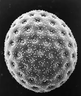
Taraxacum officinale, dandelionScanning electron microscope (SEM) image of a dandelion (x 80)
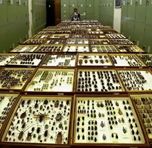
Scientist at workEntomologist studying beetle specimens at the Natural History Museum, London
A bryozoan colonyScanning electron microscope image displayed on the glass screens in the Darwin Centre, at the Natural History Museum, London
Browallia speciosa, amethystA pollen grain of the Browallia speciosa (polar view) from the family Solanaceae, the tomato family
Papilio machaon, old world swallowtailSEM image of a Papilio machaon wing
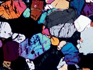
Microscope image of the Johnstown diogenite. Diogenites are coarse grained and composed primarily of one mineral, pyroxene. Field of view is 2.5mm across
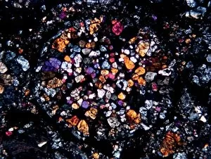
Microscope image of the Zagami shergottite. The fractures in the pyroxene mineral grains and the paler patches of glass show that the rock has been shocked. Field of view is 5mm
Microscope image of the Lodran meteorite. This meteorite is the type specimen of the Lodranite meteorites. The lodranites are related to the acaplucoites but are more course-grained
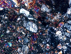
Optical microscope image of the Barwell (Type 6) chondrite. This meteorite has experienced a significant amount of heating
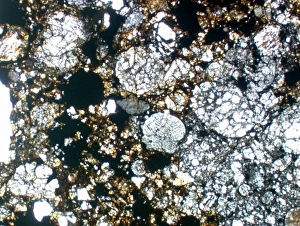
Optical microscope image of the Parnallee (Type 3) chondriteAn optical microscope image of the Parnallee (Type 3) chondrite that has experienced little heating. The chondrules are clear and well-defined. The field of view is 5mm
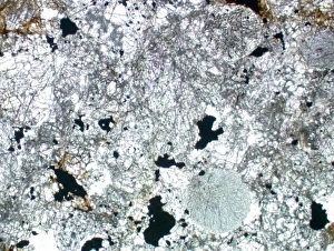
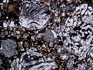
Textures of different chondrule types in the Etihudna (L4) ordinary chondrite (field of view 4mm)
Porphyritic olivine and pyroxene chondruleMicroscopic image of a porphyritic olivine and pyroxene chondrule from the Palmyra (L3) ordinary chondrite (the chondrule is about 1.8mm across)
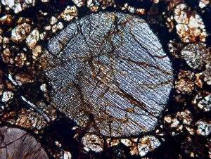
Radial pyroxene chondruleMicroscope image of a radial pyroxene chondrule from the ALH 88036 (H3.4) ordinary chondrite. The chondrule is about 2mm across
Pollen on beeScanning electron microscope (SEM) image of pollen on a bee. If the plant depends on animals for pollination, the pollen will be relatively large and sticky
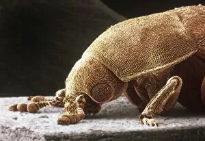
Dermestes lardarius, larder beetleScanning electron microscope image of a larder beetle (x22). These beetles are important for the damage they do, mainly through feeding on animal matter. Coloured artificially by computer
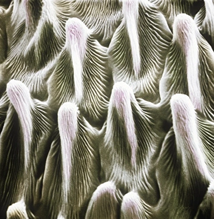
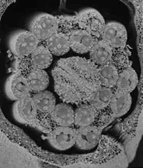
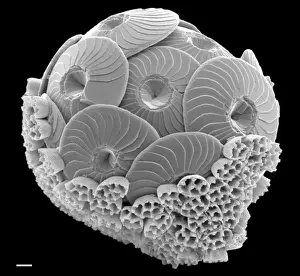
Calcidiscus leptoporus and Syracolithus quadriperforatus, coIn this scanning electron micrograph, the transition of a life-cycle stage in Calcidiscus is shown from the outer cover to the inner layer. Specimen taken from W. Mediterranean
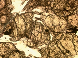
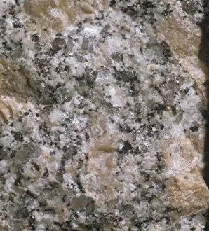
Observing structure of rockLooking at fresh granite under a microscope to study structure, granular composition can clearly be seen
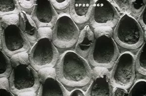
Wilbertopora woodwardi (Brydone), bryozoanScanning electron micrograph of a fossil cheilostome bryozoan. Specimen originates from the Upper Cretaceous Chalk, West Mean Station, Hampshire, U.K
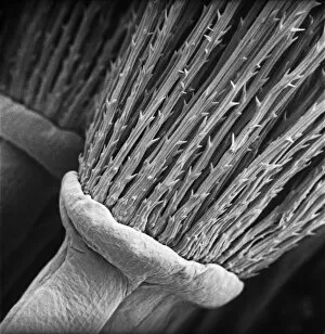
Pinus sylvestris, scots pineScanning electron microscope (SEM) image showing a pollen grain from a scots pine. Note the air bladders that help it to float through the air (x 1500 on a standard 9 cm wide print)
Chenopodium album, goosefootScanning electron microscope image of a pollen grain from a member of the goosefoot family (x 3000 on a standard 9 cm wide print)
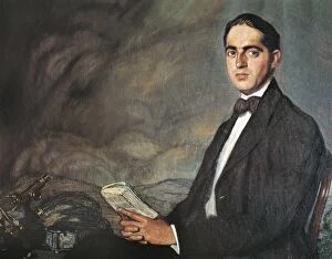
MARAяN, Gregorio (1887-1960). Spanish doctorMARAя N, Gregorio (1887-1960). Spanish doctor and writer. Portrait from 1919
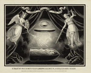
The bullet, with which our martyr President A. Lincoln was assassinated by J.W. Booth, as seen under a microscope. Date c1867. The bullet, with which our martyr President A
German Medical StudentsA group of seated (mostly female) medical students, watching a microscopic projector of what appears to be an insect. Date: 1930s
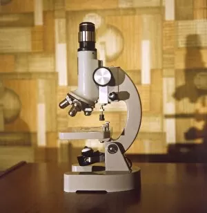
MICROSCOPEA modern microscope, in common use in school Science classrooms. Date: 1970s
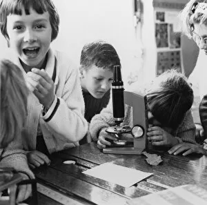
Science LessonChildren having fun with a microscope during a Science lesson at Nuffield Junior School, London. Date: 1960s
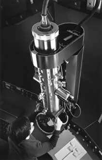
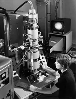
Electron MicroscopeThe EM 300 transmission electron microscope, which is used to study tiny voids or dislocations in materials or very small crystallites, A.E.R.E. Harwell. Date: October 1971
Chemical TechnologyA JEM 7A microscope can examine gas or solid interface reactions at temperatures of up to 1200oC, Chemistry Division, Harwell. Date: 1960s
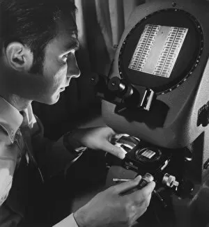
Microcircuit DevelopmentA metallurgist examines a microcircuit on the enlarging screen of a microscope at A.W.R.E. Aldermaston. Date: early 1970s
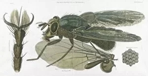
BLUEBOTTLEThe Bluebottle, or Blow-fly (MUSCA VOMITORIA) as seen through the microscope. Date: 1823
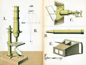
Microscope 1882Diagrams of a microscope and its constituent parts. Date: 1882
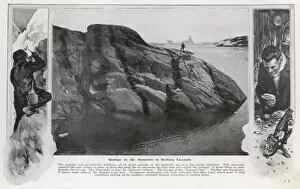
Geology in the AntarcticA photograph from Charcots " The Voyage of the Pourquoi Pas" showing a striking example of bare rock in the Graham Land area of the Antarctic
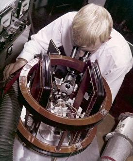
A technician doing miniature workA technician uses an unidentified instrument, incorporating a microscope and an electomagnetic circuit to work on very tiny electronic components. Photograph by Heinz Zinram