mail_outline sales@mediastorehouse.com
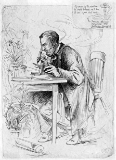
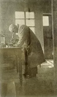
Alexander DicksonALEXANDER DICKSON botanist, at work with his microscope : A primrose by the rivers brim, no simple primrose was to him, it was a good deal more. Date: 1836 - 1887
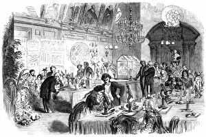
Scientific ConversazioneThe Society of Apothecaries holds a Scientific conversazione at their council chamber, Apothecaries Hall, in Blackfriars, London. The evening focused on the use of the microscope. Date: 11 April 1855
Jean Rostand / PhotoJEAN ROSTAND French biologist and writer Date: 1894 - 1977
Lumiere / Louis / Ils PhotoLOUIS-JEAN LUMIERE French chemist, industrialist and pioneer (with his brother) of cinematography Date: 1864 - 1948
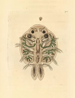
Water flea, Cyclops cyprinaceus.. Illustration drawn and engraved by Richard Polydore Nodder. Handcolored copperplate engraving from George Shaw and Frederick Nodders The Naturalists Miscellany
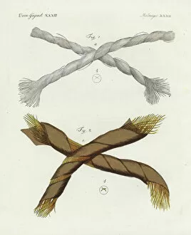
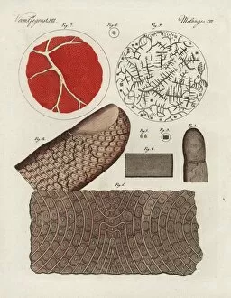
Threads of Dutch linen and gold under a microscope.. Threads of Dutch linen 1 and gold 2 magnified under a microscope. Handcolored copperplate engraving from Friedrich Bertuchs Bilderbuch fur Kinder
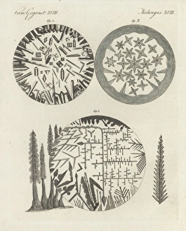
Crystals of silver solution and Dianas tree, camphor, and niter.. Crystals magnified under a microscope- silver solution and Dianas tree 1, camphor 2, and niter 3
Human finger, skin, blood, serum and salt crystals.. Human finger 1, magnified 2, epidermis 3, magnified 4, scales 5, magnified skin 6, and human blood 7, serum 8
Spermatazoa of rabbit, dog, goat and rooster.. Handcolored engraving on steel by Annedouche after a drawing by Edouard Travies from Richards New Edition of the Complete Works of Comte de Buffon
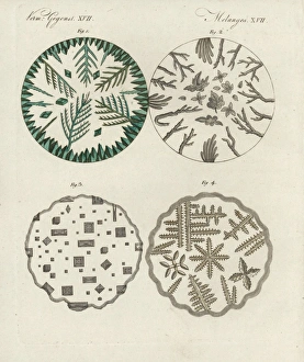
Crystals of verdigris, boric acid, common salt, and sal ammoniac.. Crystals of verdigris 1, boric acid 2, common salt 3, and sal ammoniac 4 under the microscope
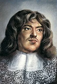
LEEUWENHOEK, Antony van (1632-1723). Dutch naturalist and optician. First to observe bacteriae. He is commonly known as the Father of Microbiology, and considered to be the first microbiologist. Oil
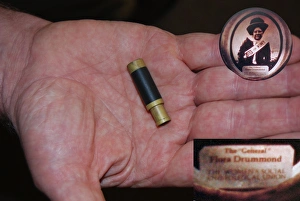
Suffragette Flora Drummond Stanhope ToyA Stanhope or optical toy, a device which allows the viewing of microphotographs without using a microscope. The stanhope was invented in 1857 by Rene Dagron, a French photographer
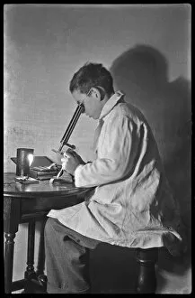
Boy with microscopeA boy in white overalls, sitting at a table examining something through a microscope. Date: 20th century
Plague scientist in ChinaWestern doctor or scientist in China during the bubonic plague outbreak; died during research work. Date: 1911
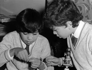
Boys with MicroscopeTwo schoolboys with a microscope in a classroom laboratory. Date: early 1970s
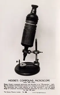
Robert Hookes MicroscopeFull-size copy of Robert Hookes Compound Microscope - held at the Science Museum, London. Hooke, an English natural philosopher, architect and polymath (1635-1703) - author of Micrographia (1665)
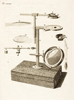
Aquatic microscope. Engraving from John Ellis, Essai sur l histoire naturelle des corallines, et d autres productions marines. Plate XXXIX. Date: 1756
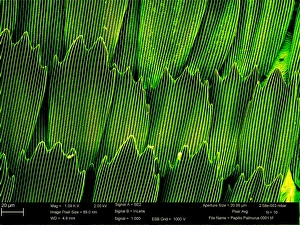
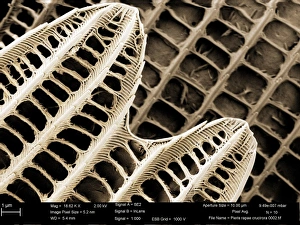
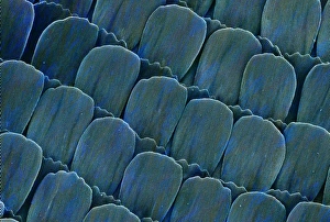
Papilio palinurus, emerald swallowtailSEM image of an emerald swallowtails wing
Pieris rapae, small whiteSEM image of the wing of a small white butterfly
Heliconius doris, doris longwingSEM image of Heliconius doris wing
Dinosaur eggshellScanning electron microscope image on display in the Darwin Centre
Ventral surface of a mite from the prostigmatic speciesScanning electron microscope image displayed on the glass screens in the Darwin Centre, at the Natural History Museum, London
Difflugia CoronaFreshwater Testate Amoebae. Magnification x 450
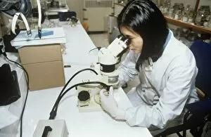
Volunteer working at the Natural History Museum, LondonVolunteer using microscope to examine zoological specimen
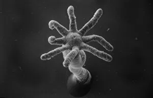
Diplopoda sp. plate millipedeScanning electron microscope image of a lateral view of the head of a plate millipede. Image displayed on the glass screens in the Darwin Centre, at the Natural History Museum, London
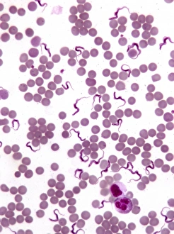
TrypanosomesScanning electron microscope image showing a trypanosoma blood smear. They have proved to be of great interest as they have evolved very differently to other better studied organisms
Taxonomic research in the fish sectionResearch on the fish collections at the Natural History Museum, London
Hydra spScanning electron microscope (SEM) image showing the stinging tentacles and mouth of the coelenterate Hydra (x 36 on a standard 9cm wide print)
Atta cethalotes, leaf-cutter antScanning electron microscope image of a leaf-cutter ant displayed in the Darwin Centre, at the Natural History Museum, London
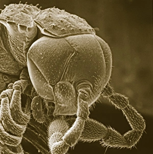
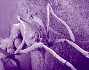
Small fly, species unknownScanning electron microscope (SEM) of a fly head. Image displayed on the glass screens in the Darwin Centre, at the Natural History Museum, London
Amphitetras, diatomScanning electron microscope (SEM) image showing the diatom Amphitetras with its ornate silica shell (x5000 on a standard 9 cm wide print). Coloured artificially by computer
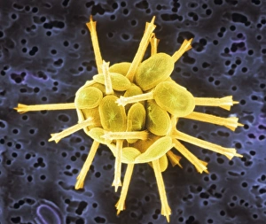
Leptoglossis ferreyraeiA pollen grain of Leptoglossis ferreyraei (polar view) from the family Solanaceae, the tomato family
Leptoglossis lomanaA pollen grain of the Leptoglossis lomana (polar view) from the family Solanacea, the tomato family
Gold in unspecified mineralScanning electron microscope image of an elemental map showing the distribution of gold (Au) in mineral samples
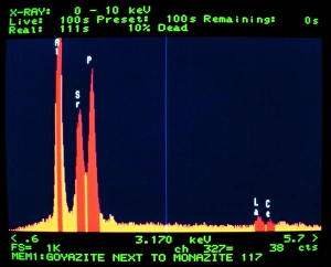
GoyaziteScanning electron microscope image of the energy-dispersive X-ray spectrum of the mineral goyazite, obtained using Link AN10000 analysis system
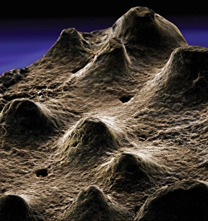
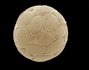
CoccolithsScanning electron microscope (SEM) image of coccoliths, these are the limestone scales surrounding the marine phytoplankton coccolithophores
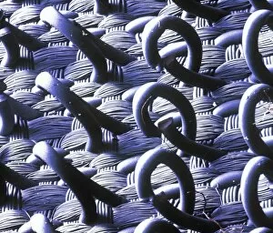
VelcroA trademarked name for a fastening tape made up of a strip of nylon with a surface of minute hooks, that fasten to another strip with a surface of uncut pile. A SEM image
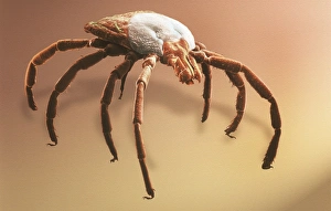
Amblyomma sp. hard backed tickScanning electron microscope view of a hard backed tick from the family Ixodidae. Coloured artificially on computer
Copper in unspecified mineralScanning electron microscope image of an elemental map showing the distribution of copper (Cu) in mineral samples
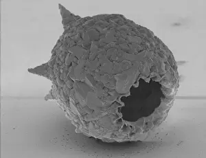
Fractured pollen grainScanning electron microscope (SEM) image showing a fractured pollen grain
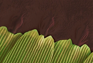
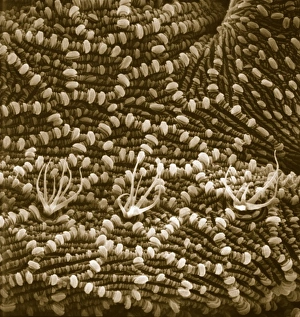
Bellis perenis, daisy petalScanning electron microscope (SEM) image of a daisy petal. Published in Close-Up (2004) by Chris Jones and Alex Ball (inside cover)
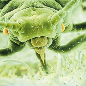
Aphis fabae, black bean aphidScanning electron microscope image showing a frontal view of a black bean aphid on leaf (x100). Aphids or plant lice are small, plant-sucking insects
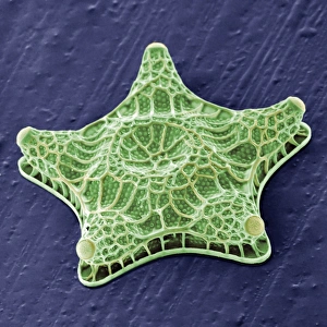
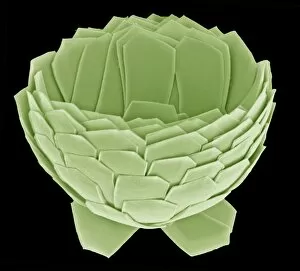
Florisphaera profundaA coccolithophore with highly modified, plate-like coccoliths. This is a very common deep dwelleing species, typically living at about 100-150m depth in the water column
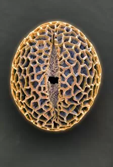
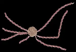
Ophiaster formosusA coccolithophore with long appendages formed of strings of highly modified coccoliths. Collected from the West Pacific. Specimen diameter 50m. False-coloured SEM image
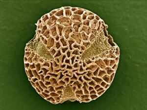
Pontosphaera japonica. A coccolithophore with relatively large, flat, coccoliths. Collected from off Hawaii. Specimen diameter 22m. False-coloured SEM image
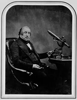
Dr James Scott Bowerbank (1797-1877)Portrait of Dr James Scott Bowerbank, an English naturalist and palaeontologist. Photographed by Maull & Polyblank, Photographers. Ca 1854
Pelargonium crispum, lemon geranium