mail_outline sales@mediastorehouse.com
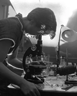
Rosalind FranklinROSALIND FRANKLIN Pioneer Molecular Biologist she made important contributions to understanding the structure of DNA
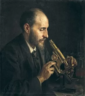
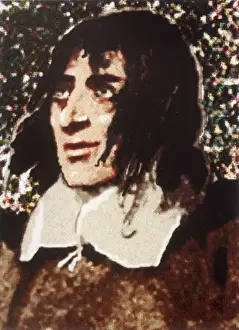
RAMON Y CAJAL, Santiago (1852-1934). Spanish
RAMON Y CAJAL, Santiago (1852-1934). Spanish doctor and histologist, Nobel Prize in 1906. Portrait in his laboratory
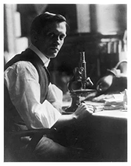
Fleming / Photo / MicroscopeSIR ALEXANDER FLEMING - Scottish bacteriologist at his desk with his microscope
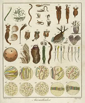
Under the Microscope / C18Animacules : microscopic creatures as seen under a microscope; the last two rows are human sperm
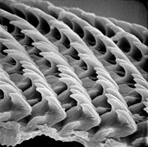
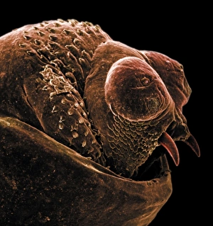
Snail teeth
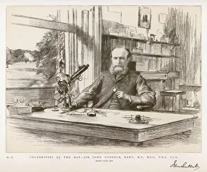
Lubbock / John / Graphic 84SIR JOHN LUBBOCK 1st BARON AVEBURY Liberal MP and writer of popular science books
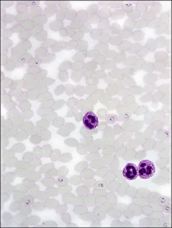
Plasmodium sp. malarial parasiteScanning electron microscope image of a malarial protozoal parasite. The parasite requires the anopheles mosquito to complete its life cycle
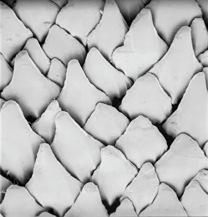
Scyliorhinus canicula, dogfishScanning electron microscope (SEM) image of the scales of a dogfish (x 40)
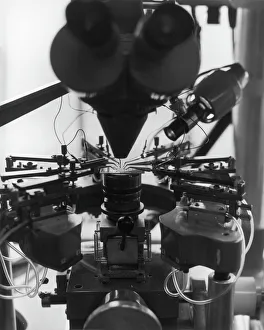
Manufacture of circuit boardsA complicated multi-component electronic device for manufacturing circuit boards. It consists of a number of miniature soldering irons to attach the electrical components and a magnifying camera
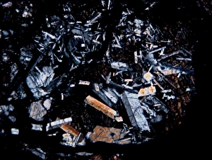
Microscope image of the Pasamonte eucriteMicroscopic image of the Pasamonte eucrite showing a basaltic texture. Field of view is 2.5mm across
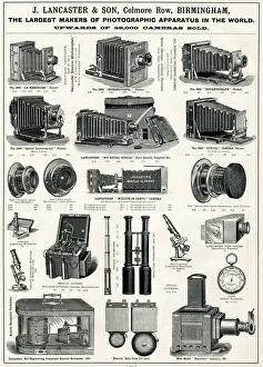
J Lancaster & Son, Colmore Row, BirminghamJ Lancaster & Son, Cameras, Photographic and Scientific Equipment, Colmore Row, Birmingham. circa 1900
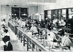
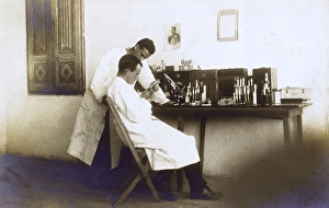
Physiology Laboratory, London School of Medicine for Women, Hunter Street. Date: 1898
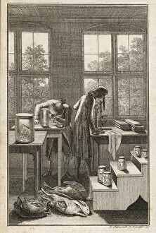
Scientists in a lab examining specimens in jars, through a microscope, with bloated animal and bird corpses on the floor. Copperplate engraving by Daniel Chodowiecki, Berlin, 1787
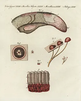
Cow tongue under the microscope: veal tongue showing different papillae 1, foliate papilla 2, filiform papillae 3, and fungiform papillae 4
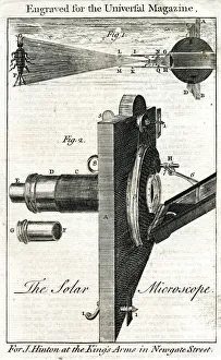
The Solar Microscope. Date: 18th century
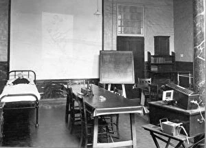
Nursing classroomAn empty classroom at the City & District Infirmary York probationer school founded in 1923. Table at centre with small microscope, and bed on left hand side with large doll. Date: circa 1923
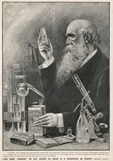
Scientist Loves CadburysAn archetypal scientist, with a flowing beard and surrounded by apparatus, studies a sample of Cadburys Cocoa; he of course concludes that it is the finest available. Date: circa 1890
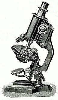
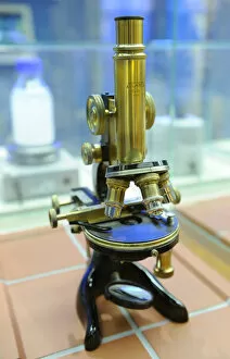
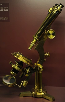
WINKELS MICROSCOPEWinkels microscope Date: late 19th century
MICROSCOPE LATE C19Date: late 19th century
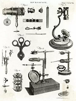
VARIOUS MICROSCOPESA selection of microscopes Date: 1797
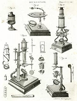
MICROSCOPE / ENC. BRITA selection of microscopes
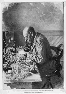
ROBERT KOCH / IN KIMBERLEYHEINRICH HERMANN ROBERT KOCH German physician and pioneer bacteriologist in search of the Rinderpest microbe at Kimberley Date: 1843 - 1910
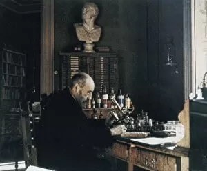
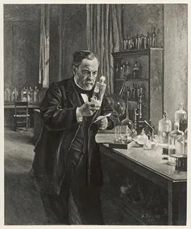
Louis Pasteur in his laboratoryLouis Pasteur (1822-1895), French chemist and microbiologist, in his laboratory. Date: 1885
The anus of a bot flyScanning electron microscope image of the anus of a bot fly. Image on display in the Darwin Centre at the Natural History Museum, London
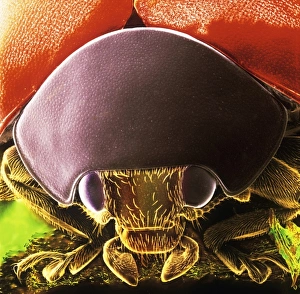
Coccinella sp. black spotted ladybirdScanning electron microscope image showing the head of a black spotted ladybird (x 9 on a standard 9 cm wide print). This image has been coloured artifically by computer
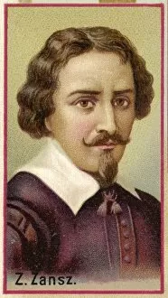
Zacharias Janssen / W InvtZACHARIAS JANSSEN or JANSEN or ZANSZ Dutch spectacle maker who invented the compound microscope
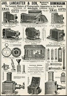
Advert for Lancaster & Sons bellows cameras 1890Advertisement for 19th century bellows cameras, pocket combination aneroid barometers, selection of scientific equipment compound microscope, achromatic microscope
Atlanta Microscope. Hartnack Berlin, 1922. Deutches Technikmuseum. Berlin. Germany
WW1 - Austrian Scientists at work in Adana, TurkeyA pair of Austrian Scientists working in secrecy in Adana, Turkey during the First Work War. Note the portrait of Archduke Franz Ferdinand
Tyrophagus casei, cheese miteScanning electron microscope image of a cheese mite (x 170). These creatures are generally considered to be a pest, however they are added to Altenburger cheese to give it flavour
Ceratodon purpureus, ceratodon moss spore capsuleScanning electron microscope (SEM) image of a ceratodon moss spore capsule (x 650 on a standard 9 cm wide print)
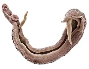
Schistosoma nasale, bloodflukeScanning electron microscope image of a parasitic bloodfluke or flatworm. Coloured artifically by computer
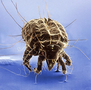
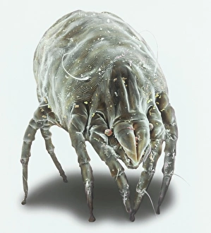
Dermatophagoides pteronyssius, dust miteScanning electron microscope image showing a dust mite (x 250 on standard 9cm wide print). This image has been artificially coloured by a computer
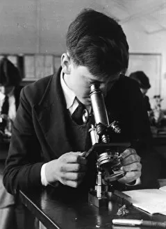
Boy Using MicroscopeA schoolboy at Lingfield Secondary uses a microscope
ROBERT HOOKE 1635 - 1703ROBERT HOOKE English scientist. Author of Micrographia (1665), in which he published results of his microscopic investigations
Robert Hooke / MicroscopeRobert Hookes microscope
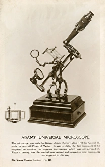
Adams Universal Microscope - made by George Adams Snr. about 1755 for King George III, while he was still Prince of Wales
Binocular microscope large Best. London, around 1890Binocular microscope large Best. Signed: R. & J. Beck Ltd London 19901. London, around 1890. The Large Best microscope was the top product of R. and J
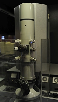
Transmission electron microscope EM9. Signed: Carl Zeiss. 1964
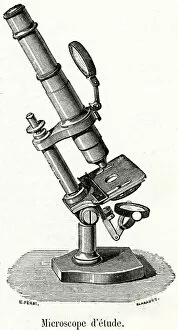
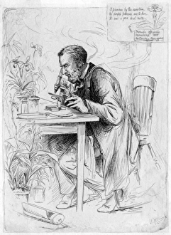
Alexander DicksonALEXANDER DICKSON botanist, at work with his microscope : A primrose by the rivers brim, no simple primrose was to him, it was a good deal more. Date: 1836 - 1887
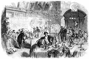
Scientific ConversazioneThe Society of Apothecaries holds a Scientific conversazione at their council chamber, Apothecaries Hall, in Blackfriars, London. The evening focused on the use of the microscope. Date: 11 April 1855
Jean Rostand / PhotoJEAN ROSTAND French biologist and writer Date: 1894 - 1977
Lumiere / Louis / Ils PhotoLOUIS-JEAN LUMIERE French chemist, industrialist and pioneer (with his brother) of cinematography Date: 1864 - 1948
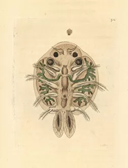
Water flea, Cyclops cyprinaceus.. Illustration drawn and engraved by Richard Polydore Nodder. Handcolored copperplate engraving from George Shaw and Frederick Nodders The Naturalists Miscellany
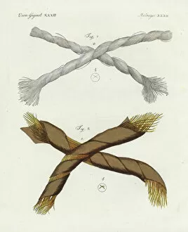
Threads of Dutch linen and gold under a microscope.. Threads of Dutch linen 1 and gold 2 magnified under a microscope. Handcolored copperplate engraving from Friedrich Bertuchs Bilderbuch fur Kinder
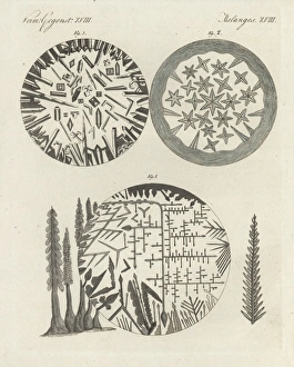
Crystals of silver solution and Dianas tree, camphor, and niter.. Crystals magnified under a microscope- silver solution and Dianas tree 1, camphor 2, and niter 3
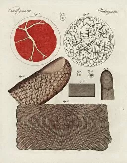
Human finger, skin, blood, serum and salt crystals.. Human finger 1, magnified 2, epidermis 3, magnified 4, scales 5, magnified skin 6, and human blood 7, serum 8