mail_outline sales@mediastorehouse.com
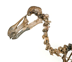
Dodo skeleton, Raphus cucullatusThe dodo is an icon of extinction, one of the first widely acknowledged cases of a species being wiped out by humans. There are so few complete dodo skeletons that we may never know exactly what they
Osteological Gallery. 5th July 1892Photograph of the Osteological Gallery. 5th July 1892. Archive ref: PH/173/2 Date: 1892
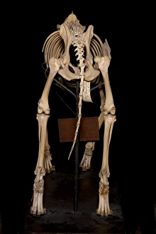
Canis sp. Eskimo Dog called Arctic KingArticulated skeleton of an Canis sp. Eskimo Dog called Arctic King
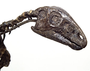
Hypsilophodon skullHypsilophodons narrow mouth would have been suitable for picking out soft shoots and leaves. Narrow mouths allow animals to select food with more care. This specimen lived 125 million years ago
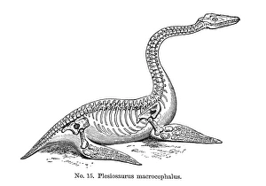
Plesiosaurus macrocephalusPlate 15 from Geology and Inhabitants of the Ancient World, by Sir Richard Owen, (1854). This marine reptile could be found during the Jurassic period between 200 and 145 million years ago. Date: 1854
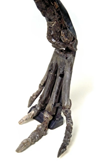
Hypsilophodon footHypsilophodons upper foot bones were long and the lower foot thin and flexible, very like todays running birds. This specimen which was discovered in England dates back 125 million years to
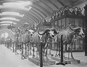
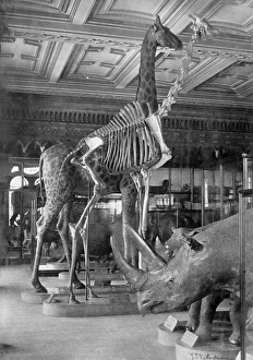
Mammal Pavilion. 5th July 1892Photograph of Giraffe and Rhinos in the Mammal Pavilion. 5th July 1892 Archive ref: PH/173/1 Date: 1892
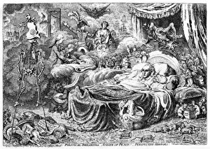
Cartoon, Political Dreamings, Visions of Peace! Perspective Horrors! by James Gillray. Showing William Windham, British War Minister, asleep and dreaming in bed, surrounded by visions
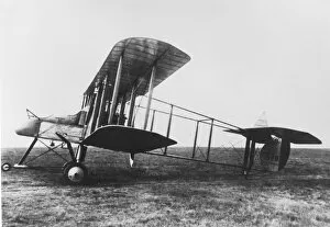
Royal Aircraft Factory FE 2b of which nearly 2, 000 were built from spring 1915. It was used as a fighter, night bomber and for reconnaissance. Serial no
Frog skeleton and edible frog, Pelophylax kl esculentusFrog skeleton and edible frog, Pelophylax kl. esculentus.. Handcolored copperplate engraving from Gottlieb Tobias Wilhelms Encyclopedia of Natural History: Amphibia, Augsburg, 1794
Loggerhead sea turtle, Caretta caretta, and skeleton of same.. Handcolored copperplate engraving from Gottlieb Tobias Wilhelms Encyclopedia of Natural History: Amphibia, Augsburg, 1794
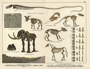
Fossil skeletons of extinct bat, giraffid, mastodon and horse.. Chromolithograph from Dr. Fr. Rolles Geology and Paleontology section in Gotthilf Heinrich von Schuberts Natural History, Schreiber
Fossil skulls and skeletons.. Chromolithograph from Dr. Fr. Rolles Geology and Paleontology section in Gotthilf Heinrich von Schuberts Natural History, Schreiber, Munich, 1886
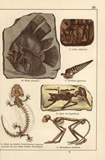
Fossil skeletons of fish, shell, bird and monkey.. Chromolithograph from Dr. Fr. Rolles Geology and Paleontology section in Gotthilf Heinrich von Schuberts Natural History, Schreiber, Munich, 1886
Fossil skeleton, tooth, skull and excrement.. Chromolithograph from Dr. Fr. Rolles Geology and Paleontology section in Gotthilf Heinrich von Schuberts Natural History, Schreiber, Munich, 1886
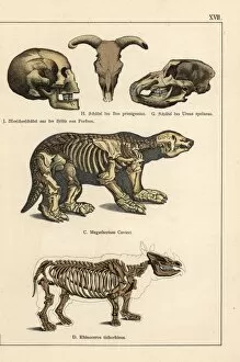
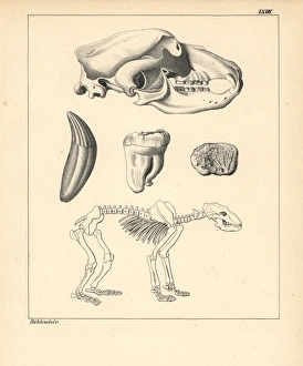
Skeleton, skull and tooth of the cave bear, Ursus spelaeus.. Handcolored lithograph from Dr. F.A. Schmidts Petrefactenbuch, published in Stuttgart, Germany, 1855 by Verlag von Krais & Hoffmann. Dr
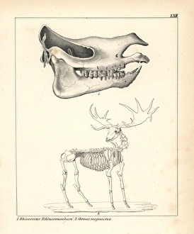
Skull of the Rhinoceros schleirmacheri and skeleton of the Irish elk, Cervus megaceros.. Handcolored lithograph from Dr. F.A
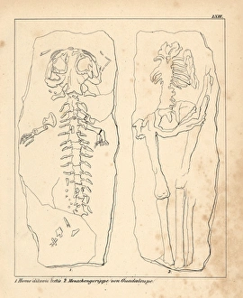
Diluvian human skeleton known as Homo diluvii testis, and a human skeleton from Guadalupe.. Handcolored lithograph from Dr. F.A
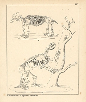
Skeleton of the American mastodon, Mammut americanum, and giant ground sloth, Mylodon robustus.. Handcolored lithograph from Dr. F.A
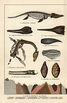
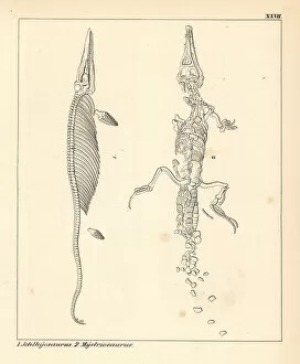
Fossil dinosaur skeletons: Ichthyosaurus and teleosaurid crocodyliform Mystriosaurus.. Handcolored lithograph from Dr. F.A
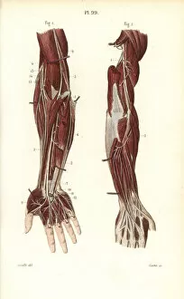
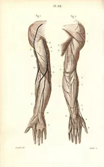
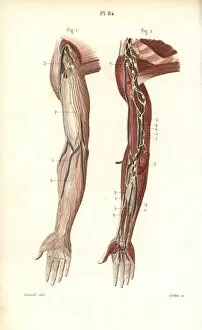
Deep nervous system to the arm and hand.. Handcolored steel engraving from Dr. Joseph Nicolas Masses Pocket Anatomy of the Human Body, Paris, 1864
Brachial plexus.. Handcolored steel engraving from Dr. Joseph Nicolas Masses Pocket Anatomy of the Human Body, Paris, 1864
Nerves to the arm and hand.. Handcolored steel engraving from Dr. Joseph Nicolas Masses Pocket Anatomy of the Human Body, Paris, 1864
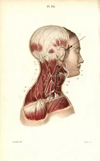
Cervical plexus.. Handcolored steel engraving from Dr. Joseph Nicolas Masses Pocket Anatomy of the Human Body, Paris, 1864
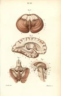
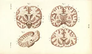
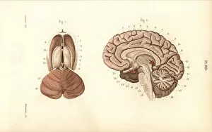
Sections through the brain, cerebellum and ventricles.. Handcolored steel engraving from Dr. Joseph Nicolas Masses Pocket Anatomy of the Human Body, Paris, 1864
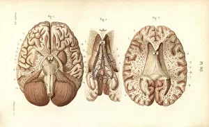
Brain, pons, medulla oblongata, pineal gland, etc.. Handcolored steel engraving from Dr. Joseph Nicolas Masses Pocket Anatomy of the Human Body, Paris, 1864
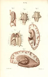
Sections through the brain and pineal gland.. Handcolored steel engraving from Dr. Joseph Nicolas Masses Pocket Anatomy of the Human Body, Paris, 1864
Sections through the brain.. Handcolored steel engraving from Dr. Joseph Nicolas Masses Pocket Anatomy of the Human Body, Paris, 1864
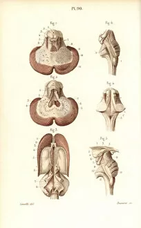
Sections through the cerebellum, ventricular system.. Handcolored steel engraving from Dr. Joseph Nicolas Masses Pocket Anatomy of the Human Body, Paris, 1864
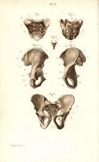
Sacrum, coccyx and pelvis bones.. Handcolored steel engraving from Dr. Joseph Nicolas Masses Pocket Anatomy of the Human Body, Paris, 1864
Cross sections through the brain.. Handcolored steel engraving from Dr. Joseph Nicolas Masses Pocket Anatomy of the Human Body, Paris, 1864
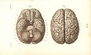
Brain from above and below.. Handcolored steel engraving from Dr. Joseph Nicolas Masses Pocket Anatomy of the Human Body, Paris, 1864
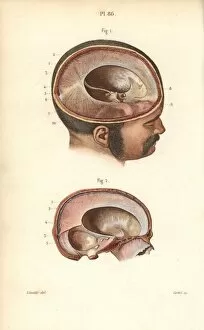
Cross sections through the skull.. Handcolored steel engraving from Dr. Joseph Nicolas Masses Pocket Anatomy of the Human Body, Paris, 1864
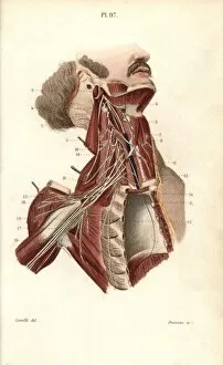
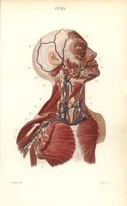
Lymph nodes and vessels in the head, neck and chest.. Handcolored steel engraving from Dr. Joseph Nicolas Masses Pocket Anatomy of the Human Body, Paris, 1864
Lymphatic system in the arm.. Handcolored steel engraving from Dr. Joseph Nicolas Masses Pocket Anatomy of the Human Body, Paris, 1864
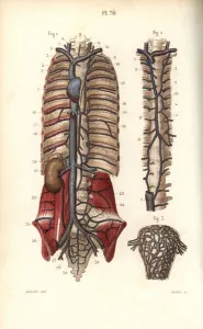
The thoracic canal.. Handcolored steel engraving from Dr. Joseph Nicolas Masses Pocket Anatomy of the Human Body, Paris, 1864
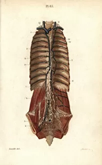
Lymphatic system to the thorax and abdomen.. Handcolored steel engraving from Dr. Joseph Nicolas Masses Pocket Anatomy of the Human Body, Paris, 1864
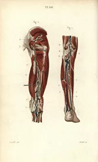
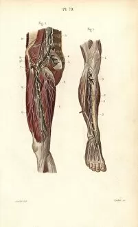
Lymph nodes and vessels deep in the back of the leg.. Handcolored steel engraving from Dr. Joseph Nicolas Masses Pocket Anatomy of the Human Body, Paris, 1864
Lymphatic system to the abdomen.. Handcolored steel engraving from Dr. Joseph Nicolas Masses Pocket Anatomy of the Human Body, Paris, 1864
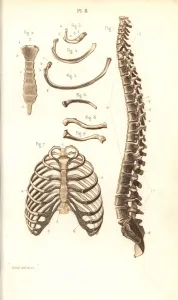
Spine, ribs, vertebrae and sternum.. Handcolored steel engraving from Dr. Joseph Nicolas Masses Pocket Anatomy of the Human Body, Paris, 1864
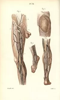
Lymph nodes and vessels deep in the leg.. Handcolored steel engraving from Dr. Joseph Nicolas Masses Pocket Anatomy of the Human Body, Paris, 1864
Lymphatic system in the leg and foot.. Handcolored steel engraving from Dr. Joseph Nicolas Masses Pocket Anatomy of the Human Body, Paris, 1864
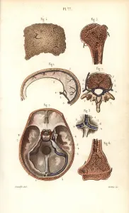
Sinuses or cavities in skull, humerus and vertebrae.. Handcolored steel engraving from Dr. Joseph Nicolas Masses Pocket Anatomy of the Human Body, Paris, 1864
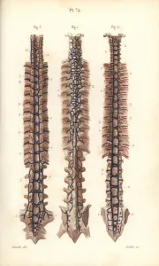
Veins to the spine.. Handcolored steel engraving from Dr. Joseph Nicolas Masses Pocket Anatomy of the Human Body, Paris, 1864
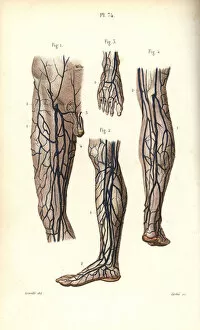
Veins to the leg and foot.. Handcolored steel engraving from Dr. Joseph Nicolas Masses Pocket Anatomy of the Human Body, Paris, 1864
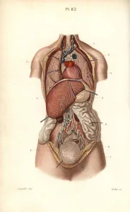
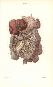
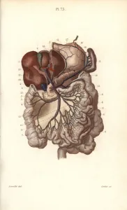
Veins to the stomach, liver and intestines.. Handcolored steel engraving from Dr. Joseph Nicolas Masses Pocket Anatomy of the Human Body, Paris, 1864
Circulatory system to the spine and uterus.. Handcolored steel engraving from Dr. Joseph Nicolas Masses Pocket Anatomy of the Human Body, Paris, 1864
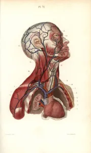
Circulatory system to the head and torso.. Handcolored steel engraving from Dr. Joseph Nicolas Masses Pocket Anatomy of the Human Body, Paris, 1864