mail_outline sales@mediastorehouse.com
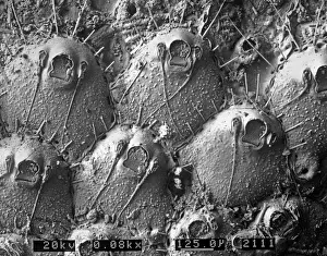
Volcanic glass, Peles hairScanning electron microscope image of a sample of volcanic glass from Mt. Pele, produced to evaluate different types of laser in Laser Ablation Inductively Coupled Plasma Mass Spectrometry
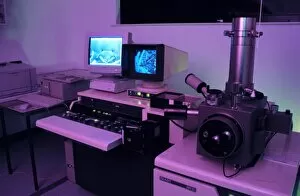
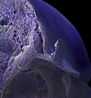
Variable pressure scanning electron microscopeThis electron microscope allows the imaging of samples without any preparation
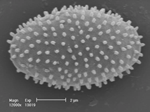
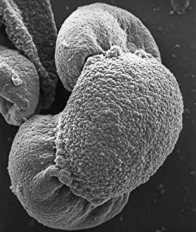
Myxomycetes, plasmodial slime mouldScanning electron microscope image of a plasmodial slime mould spore (x12000). This mould spends most of its life as a single cell; when they reproduce they form a slug-like blob that can travel
Collembola ocelli, springtailScanning electron microscope image of the springtail with simple eyes (x 1.2K)
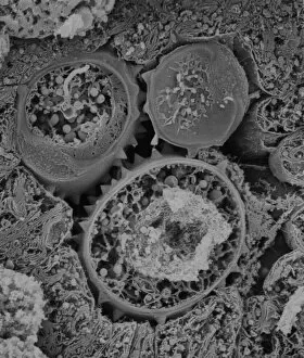
Copper in unspecified mineralScanning electron microscope image of an elemental map showing the distribution of copper (Cu) in mineral samples
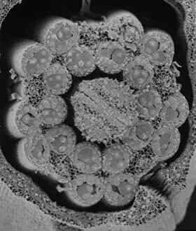
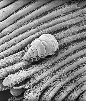
OstracodScanning electron microscope image of an ostracod, an arthropod where the body is enclosed in a carapace (x 220)
Collembola sp. springtailScanning electron microscope image of a springtail showing the characteristic pattern on the cuticle surface (x 3.5K)
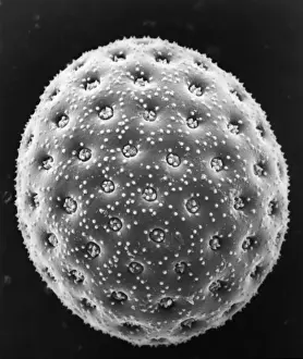
Fractured pollen grainScanning electron microscope (SEM) image showing a fractured pollen grain
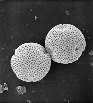
Populus nigra, lombardy or black poplar pollenScanning electron microscope image (x 1500) of black poplar pollen grains showing a characteristic granular surface ornamentation and no apertures (inaperturate)
Butterfly wing scale (part)
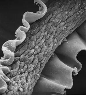
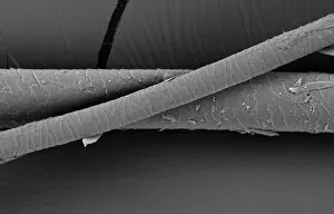
Human hairScanning electron microscope (SEM) image showing a human hair with the cuticle reflexed
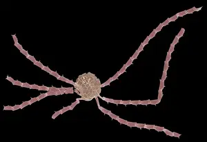
Ophiaster formosusA coccolithophore with long appendages formed of strings of highly modified coccoliths. Collected from the West Pacific. Specimen diameter 50m. False-coloured SEM image
Calliphora vicina, blowfly or blue bottleScanning electron microscope (SEM) image of a blowflys wing
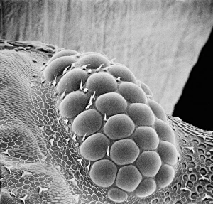
Fractured antherScanning electron microscope (SEM) image showing a fractured anther, otherwise known as the sac, which contains the pollen in the male sex organs (stamens)
Asteraceae, daisyScanning electron microscope image of the fractured surface of an anther showing a developing pollen grain from a member of the daisy or Asteraceae family ( X 3000)
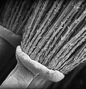
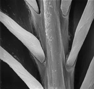
Lumbricus terrestris, earthwormScanning electron microscope (SEM) image showing the chaeta/setae - involved in the locomotion on an earthworm
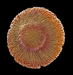
Taraxacum officinale, dandelionScanning electron microscope (SEM) image of a dandelion (x 80)
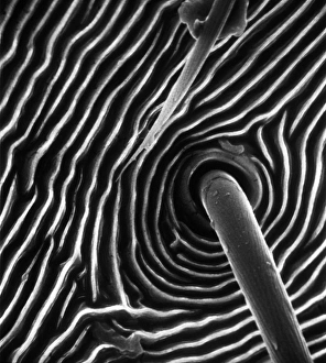
Spider trichobothrium hairScanning electron microscope (SEM) image of the base of a trichobothrium hair (x 1, 000). The hair is an air-movement sensor extending from the pit in the cuticle of a spiders leg
Fagus sylvatica, European beech pollenScanning electron microscope picture (X1500) showing a pollen grain as seen from the side. The image shows one of the three laterally-placed aperture furrows with a small pore in the centre
Fraxinus excelsior, weeping ash pollenScanning electron microscope picture (x 1500) of ash pollen grains from above, with three furrowed apertures (trizonocolporate)
Calyptrolithophora papillifera, holococcolithAn SEM of a holococcolith, a nano-fossil, with flat top
A bryozoan colonyScanning electron microscope image displayed on the glass screens in the Darwin Centre, at the Natural History Museum, London
Cystopteris diaphana, diaphanous bladder fernAn SEM showing a close-up of the spiny-lacunar surface of the diaphanous bladder fern (Cystopteris diaphana) spore. Photographed using Philips XL30 SEM
Hair of the DogA scanning electron micrograph (SEM) of a dog hair
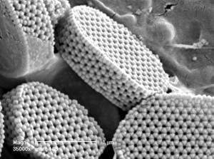
Sugar grainsA scanning electron microscope (SEM) image of sugar grains, artificially coloured by computer
Vitis sp. white grapeA scanning electron microscope (SEM) image of a white grape (Vitis sp.), artificially coloured by computer
Browallia speciosa, amethystA pollen grain of the Browallia speciosa (polar view) from the family Solanaceae, the tomato family
Vaccinium sp. blueberryA scanning electron microscope (SEM) image of a blueberry (Vaccinium sp.), artificially coloured by computer
Solanum sp. tomatoA scanning electron microscope (SEM) image of a tomato (Solanum sp.), artificially coloured by computer
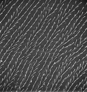
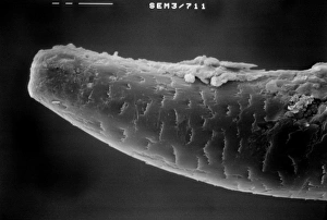
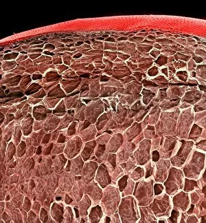
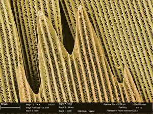
Isurus oxyrinchus, mako sharkScanning Electron Microscope image of mako shark skin
Porcellio sccaber, woodlouseScanning electron microscope (SEM) image showing all the units that make up the compound eye of a woodlouse
Papilio machaon, old world swallowtailSEM image of a Papilio machaon wing
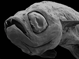
Danionella dracula, minnowSEM image of the Danionella dracula. This tiny 17mm fish has evolved many unique and unusual characteristics, the most spectacular of which are jaw modifications that resemble true teeth
Feather detail
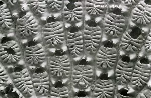
Actinopora disticha, bryozoanScanning electron micrograph of a fossil cyclostome bryozoan from the Cretaceous Chalk, Santonian, Kent
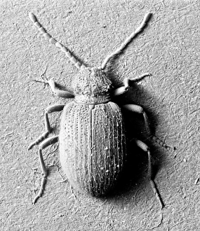
Ptinus tectus, spider beetleScanning electron microscope image of a spider beetle (x 9). The long antennae, hairy body and waist-like constriction give this beetle the appearance of a spider
Aspidelectra melolontha, bryozoanScanning electron micrograph. Zooids of a bleached colony of a modern cheilostome bryozoan. A recent specimen from Sheppey, Kent
Pinus sylvestris, scots pineScanning electron microscope (SEM) image showing a pollen grain from a scots pine. Note the air bladders that help it to float through the air (x 1500 on a standard 9 cm wide print)
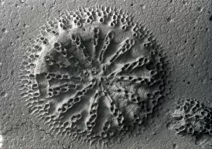
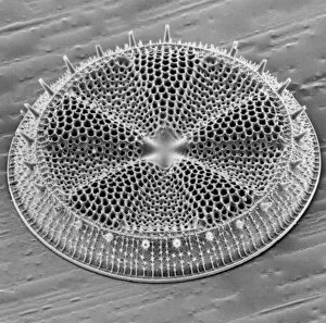
Actinoptychus, diatomScanning electron microscope image of the exterior valve of the diatom Actinoptychus (x 500 on a standard 9 cm wide print)
Chenopodium album, goosefootScanning electron microscope image of a pollen grain from a member of the goosefoot family (x 3000 on a standard 9 cm wide print)
YAхZ DE LA ALMEDINA, Fernando (1489-1536);Gassendi" YAх Z DE LA ALMEDINA, Fernando (1489-1536); Gassendi, Pierre (1592-1665); SEM, Georges Goursat, called (1863-1934); BEARDSLEY, Aubrey Vincent (1872-1898); FIRENZE
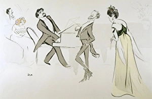
Reynaldo Hahn / Sem 14 / 2REYNALDO HAHN French musician performing chez Madeleine Lemaire (the model for Prousts Mme Verdurin) in 1904 Date: 1874 - 1947
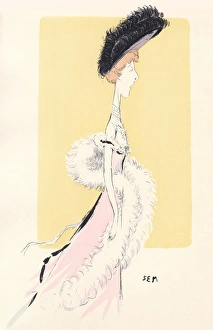
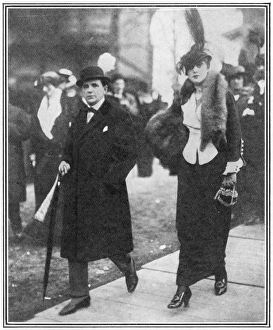
Costume / Sem 1901Depicted in caricature actress Liane de Pougy illustrates the current silhouette with extra height suggested by the plumed picture hat & trained skirt. A white feather boa is also worn Date: 1901
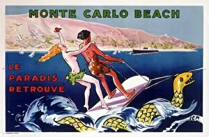
Poster for Monte-Carlo Beach, Monaco, printed by Draeger. Paradise Found
The Cartoonist SemThe cartoonist and caricaturist, Georges Goursat, otherwise known as " Sem", (1863 - 1934), pictured at the races at Auteuil with Madame Roxane