mail_outline sales@mediastorehouse.com
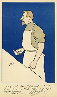
Eugene-Louis Doyen / SemEUGENE-LOUIS DOYEN French medical Date: 1859 - 1916
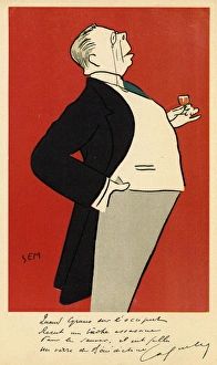
Coquelin(A) / Sem BenedictCONSTANT COQUELIN Aine (elder) French actor enjoying a small glass of wine Date: 1841 - 1909
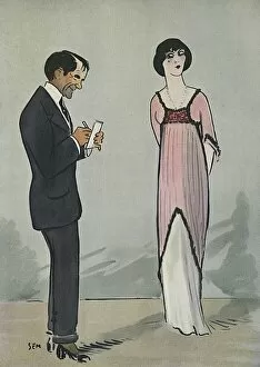
Eva Lavallerie, French actress, being sketched by Sem Date: 1910
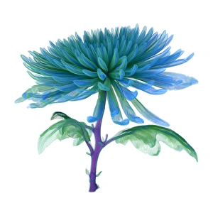
Chrysanthemum, CT scan imageCT Scan image of a Chrysanthemum
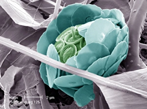
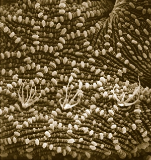
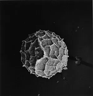
Syracosphaera anthosCoccosphere from the Western Mediterranean. False coloured to show the shell is formed of inner and outer layers of coccoliths with very different structure
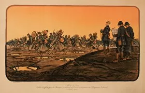
WWI Poster, French War LoanDesign by Sem, French War Loan poster, October 1918. Showing soldiers on the battlefield. Date: 1918
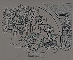
Paris: Le Mot 1914-1915 Bound folioParis: Le Mot. 1914-1915. Bound folio with a book cover by A.G. Gonon and a book plate for H.P. Gassier (French cartoonist - 1883-1951)
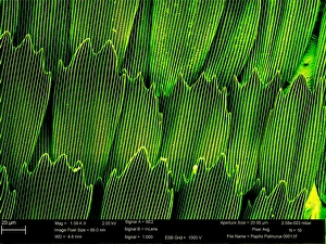
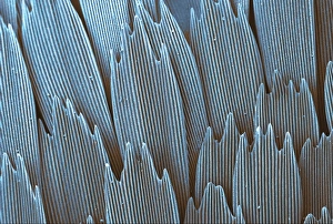
Papilio palinurus, emerald swallowtailSEM image of an emerald swallowtails wing
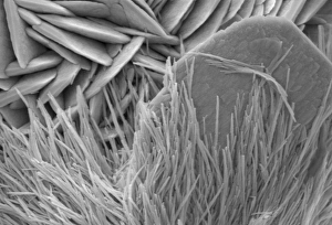
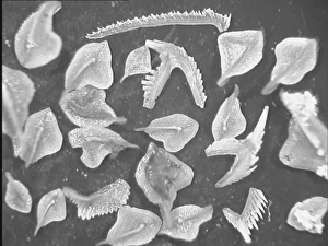
Pieris rapae, small whiteSEM image of the wing of a small white butterfly
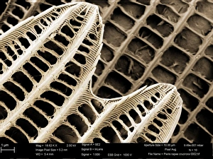
Papilio machaon, old world swallowtailSEM image of Papilio machaon wing
Malachite comprises of (copper carbonate hydroxide). Malachite has distinctive green banding and belongs to the carbonate class
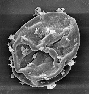
Scyphosphaera apsteinii. SEM image of an equatorial coccolith
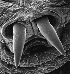
Ventral surface of a mite from the prostigmatic speciesScanning electron microscope image displayed on the glass screens in the Darwin Centre, at the Natural History Museum, London
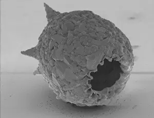
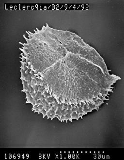
Visbyshaera oligofurcata, acritarchScanning electron microscope image of a microfossil belonging to a group of marine phytoplanktonic organisms known as acritarchs that teemed in Silurian seas about 415 Ma ago
Oestridae, botfly larvaScanning electron microscope image of a botfly larva. They are parasites feeding on skin in the case of warble flies, nostrils in the flies that affect sheep and deer
Difflugia CoronaFreshwater Testate Amoebae. Magnification x 450
Lycopod
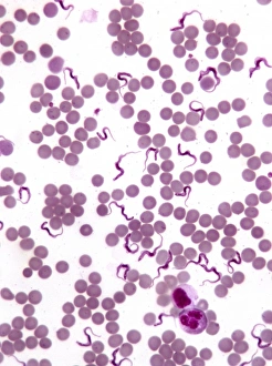
TrypanosomesScanning electron microscope image showing a trypanosoma blood smear. They have proved to be of great interest as they have evolved very differently to other better studied organisms
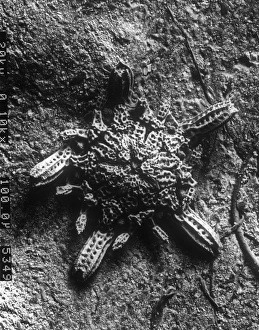
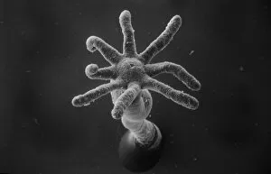
Ophioctenella sp. brittle starScanning electron microscope image of the post-larval stage of a brittle star (x 110) A newly described species 1994
Rusty screw
Surface of a rusty screw
MatchstickScanning electron microscope (SEM) image showing the fractured surface of a matchstick (x 400 on a standard 9 cm wide print)
Selaginella kraussiana, spikemossScanning electron microscope image of the female spore of Krauss spikemoss (x 150 on a standard 9 cm wide print)
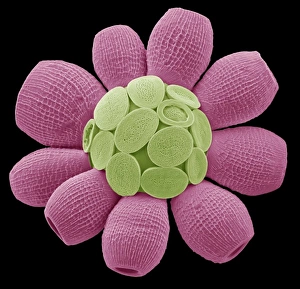
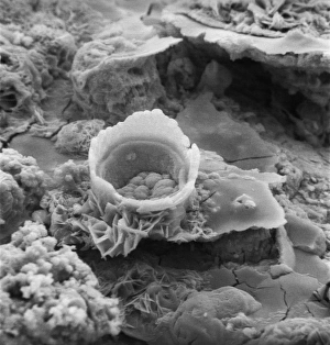
Hydra spScanning electron microscope (SEM) image showing the stinging tentacles and mouth of the coelenterate Hydra (x 36 on a standard 9cm wide print)
Conodont fossilsScanning electron microscope image of fossils from the Devonian period of northern Estonia, about 465 Ma old ( x 4.2). These creatures are still a mystery to paleontologists
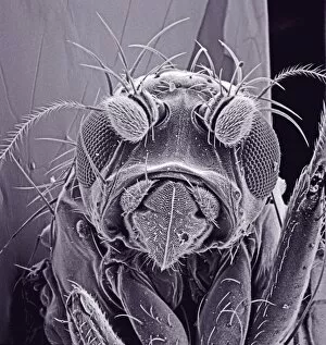
Small fly, species unknownScanning electron microscope (SEM) of a fly head. Image displayed on the glass screens in the Darwin Centre, at the Natural History Museum, London