mail_outline sales@mediastorehouse.com
Lycopod
Neanura ?muscorum, plant mouldA magnifiied image of oak leaf mould. Specimen originates from Horstead, Norwich
Rusty screw
Surface of a rusty screw
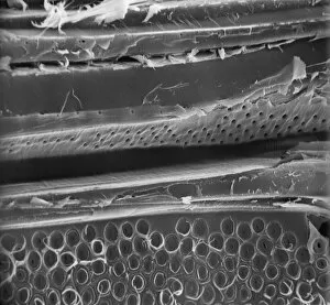
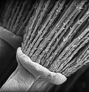
MatchstickScanning electron microscope (SEM) image showing the fractured surface of a matchstick (x 400 on a standard 9 cm wide print)
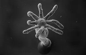
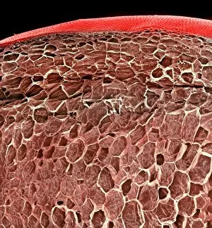
Hydra spScanning electron microscope (SEM) image showing the stinging tentacles and mouth of the coelenterate Hydra (x 36 on a standard 9cm wide print)
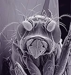
Small fly, species unknownScanning electron microscope (SEM) of a fly head. Image displayed on the glass screens in the Darwin Centre, at the Natural History Museum, London
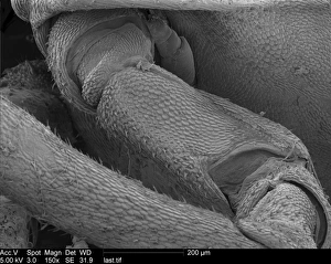
Woodlouse antennaScanning Electron Microscope (SEM) image of woodlouse antenna
OatsA scanning electron microscope (SEM) image of oats, artificially coloured by computer
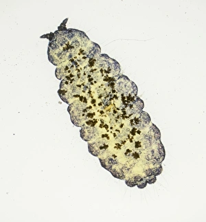
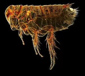
Hystrichopsylla talpae talpae, mole fleaA macro photograph of the largest flea in the UK, the mole flea (Hystrichopsylla talpae talpae), which is common on small mammals throughout the UK
Spinacia oleracea, spinachA scanning electron microscope (SEM) image of spianch (Spinacia oleracea), artificially coloured by computer
Solanum sp. tomatoA scanning electron microscope (SEM) image of a tomato (Solanum sp.), artificially coloured by computer
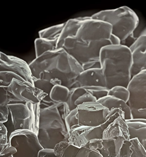
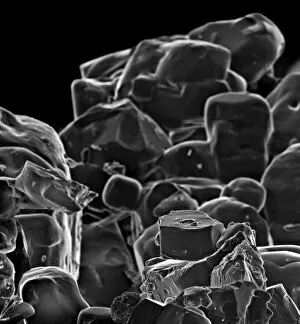
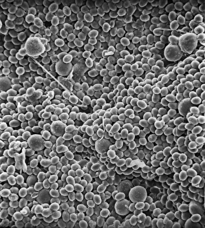
Table saltA scanning electron microscope (SEM) image of table salt, artificially coloured by computer
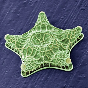
Amphitetras, diatomScanning electron microscope (SEM) image showing the diatom Amphitetras with its ornate silica shell (x5000 on a standard 9 cm wide print). Coloured artificially by computer
Vitis sp. red grapeA scanning electron microscope (SEM) image of a red grape (Vitis sp.), artificially coloured by computer
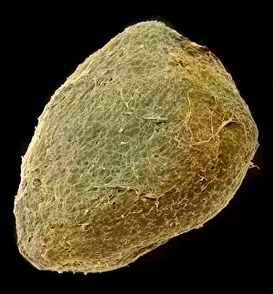
Solanum sp. tomato seedA scanning electron microscope (SEM) image of a tomato seed (Solanum sp.), artificially coloured by computer
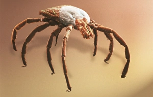
Amblyomma sp. hard backed tickScanning electron microscope view of a hard backed tick from the family Ixodidae. Coloured artificially on computer
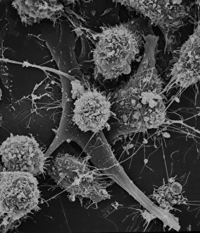
Cells on glassScanning electron microscope (SEM) image of cells on glass (x 2K)
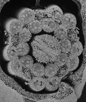
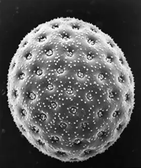
Fractured pollen grainScanning electron microscope (SEM) image showing a fractured pollen grain
Pelargonium crispum, lemon geranium
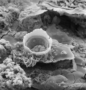
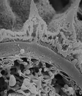
Fractured antherScanning electron microscope (SEM) image showing a fractured anther, otherwise known as the sac, which contains the pollen in the male sex organs (stamens)
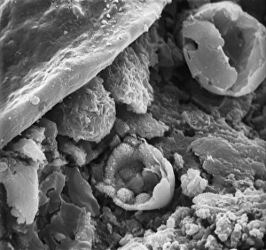
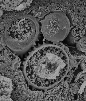
Asteraceae, daisyScanning electron microscope image of the fractured surface of an anther showing a developing pollen grain from a member of the daisy or Asteraceae family ( X 3000)
Taraxacum officinale, dandelionScanning electron microscope (SEM) image of a dandelion (x 80)
Fragaria sp. strawberryA scanning electron microscope (SEM) image of a strawberry (Fragaria sp.), artificially coloured by computer
Sugar grainsA scanning electron microscope (SEM) image of sugar grains, artificially coloured by computer
Vitis sp. white grapeA scanning electron microscope (SEM) image of a white grape (Vitis sp.), artificially coloured by computer
Vaccinium sp. blueberryA scanning electron microscope (SEM) image of a blueberry (Vaccinium sp.), artificially coloured by computer
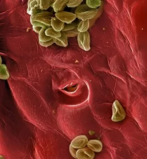
Pollen on beeScanning electron microscope (SEM) image of pollen on a bee. If the plant depends on animals for pollination, the pollen will be relatively large and sticky
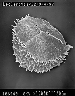
Chenopodium album, goosefootScanning electron microscope image of a pollen grain from a member of the goosefoot family (x 3000 on a standard 9 cm wide print)
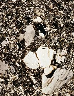
Granite from Ailsa CraigA photomicrograph of granite taken between crossed polarisers. Granite is an igneous rock
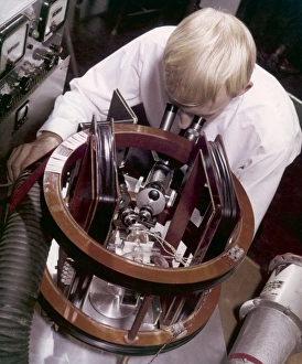
A technician doing miniature workA technician uses an unidentified instrument, incorporating a microscope and an electomagnetic circuit to work on very tiny electronic components. Photograph by Heinz Zinram
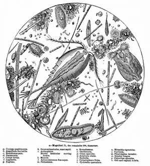
The Water of the Serpentine Magnified 200 Times, 1857Engraving showing a sample of water from the Serpentine, Hyde Park, London viewed at 200 times magnification, 1857. This particular sample was taken from the upper portion of the Serpentine
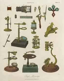
Selection of MicroscopesA selection of single microscopes
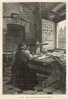
Galileo / Telescope / StudyGalileo calculates the magnification of his telescope