mail_outline sales@mediastorehouse.com
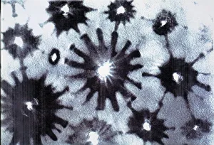
Crysotile asbestosScanning electron micrograph of 5-Fold symmetry in crysotile asbestos. Magnification on the 5 x4 transparency = X 600, 000
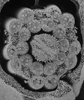
LiverScanning electron microscope (SEM) image of a section through a liver (x 7000), an organ that has over 500 functions in the human body (x 800)
Human cellIllustration of a highly magnified section through a human cell. Page 8 from Human Biology, 1977
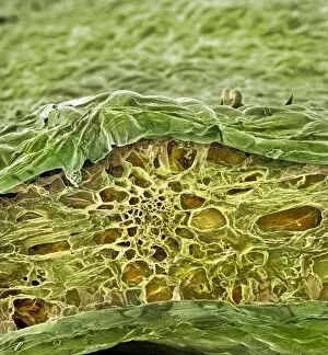
Taraxacum officinale, dandelion (fruiting head)Scanning electron microscope image showing a vertical section through an unripe fruiting head of a dandelion in the yellow flower stage. Colour added artificially by computer
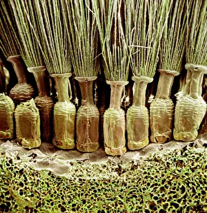
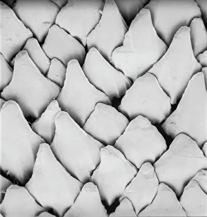
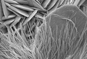
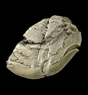
Snail teeth
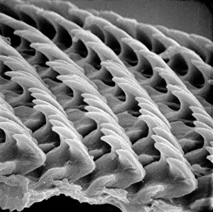
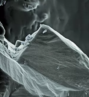
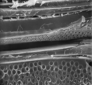
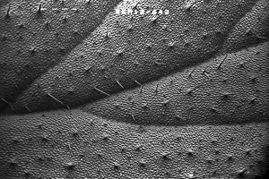
Scyliorhinus canicula, dogfishScanning electron microscope (SEM) image of the scales of a dogfish (x 40)
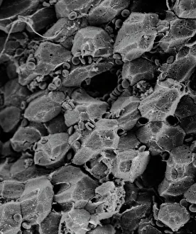
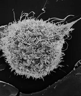
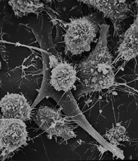
T2 cell cultureScanning electron microscope image showing a T2 cell culture (x 4K)
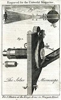
The Solar Microscope. Date: 18th century
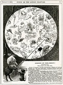
Cartoon, Essence of Parliament (MPs)Cartoon, Essence of Parliament -- Mr Punch shows members of the House of Commons as microscopic bugs in a petri dish. 1883
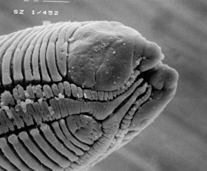
RoundwormScanning electron microscope (SEM) image of a parasitic roundworms head (x 1000 on a standard 9 cm wide print)
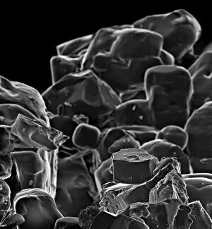
Sea saltA scanning electron microscope (SEM) image of sea salt, artificially coloured by computer
Fragaria sp. strawberryA scanning electron microscope (SEM) image of a strawberry (Fragaria sp.), artificially coloured by computer
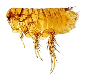
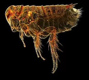
Hystrichopsylla talpae talpae, mole fleaA macro photograph of the largest flea in the UK, the mole flea (Hystrichopsylla talpae talpae), which is common on small mammals throughout the UK
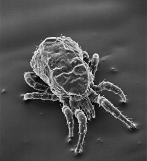
Dermanyssus gallinae, red or poultry miteScanning electron microscope image of the red or poutry mite. Adults appear red when engorged with blood, but otherwise are black, grey or white. Females are about 1mm long
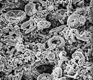
CoccolithScanning electron microscope (SEM) image of a Folkestone chalk surface with Cretaceous coccoliths (x2500 on a standard 9 cm wide print)
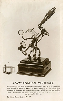
Adams Universal Microscope - made by George Adams Snr. about 1755 for King George III, while he was still Prince of Wales
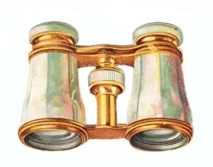
Christmas card in the shape of a pair of binoculars or opera glasses. (2 of 2) Date: circa 1890s
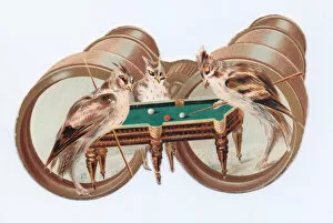
Greetings card in the shape of a pair of binoculars, with three humanised birds playing snooker in the foreground. Date: circa 1890s
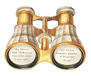
New Year card in the shape of a pair of binoculars or opera glasses. circa 1890s
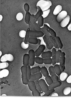
Bacillus sp. bacteriaBacteria are the most diverse and ubiquitous soil organisms present on Earth
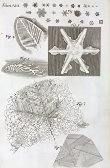
Schem VIII from Robert Hookes MicrographiaSchem VIII located between pages 88 & 89 in Micrographia: or Some physical descriptions of minute bodies made by magnifying glasses, with observations and enquiries thereupon
Malachite comprises of (copper carbonate hydroxide). Malachite has distinctive green banding and belongs to the carbonate class
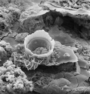
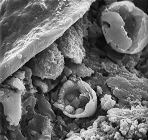
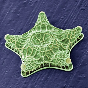
Difflugia CoronaFreshwater Testate Amoebae. Magnification x 450
Lycopod
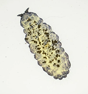
Neanura ?muscorum, plant mouldA magnifiied image of oak leaf mould. Specimen originates from Horstead, Norwich
Rusty screw
Surface of a rusty screw
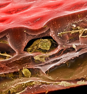
MatchstickScanning electron microscope (SEM) image showing the fractured surface of a matchstick (x 400 on a standard 9 cm wide print)
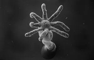
Hydra spScanning electron microscope (SEM) image showing the stinging tentacles and mouth of the coelenterate Hydra (x 36 on a standard 9cm wide print)
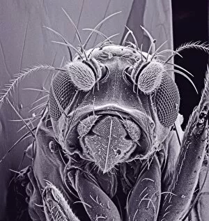
Small fly, species unknownScanning electron microscope (SEM) of a fly head. Image displayed on the glass screens in the Darwin Centre, at the Natural History Museum, London
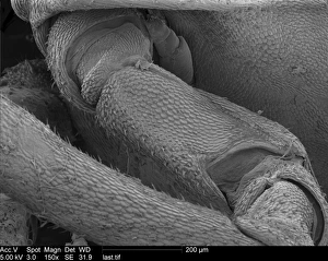
Woodlouse antennaScanning Electron Microscope (SEM) image of woodlouse antenna
OatsA scanning electron microscope (SEM) image of oats, artificially coloured by computer
Spinacia oleracea, spinachA scanning electron microscope (SEM) image of spianch (Spinacia oleracea), artificially coloured by computer
Solanum sp. tomatoA scanning electron microscope (SEM) image of a tomato (Solanum sp.), artificially coloured by computer
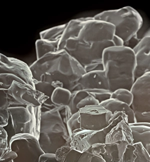
Table saltA scanning electron microscope (SEM) image of table salt, artificially coloured by computer
Amphitetras, diatomScanning electron microscope (SEM) image showing the diatom Amphitetras with its ornate silica shell (x5000 on a standard 9 cm wide print). Coloured artificially by computer
Vitis sp. red grapeA scanning electron microscope (SEM) image of a red grape (Vitis sp.), artificially coloured by computer
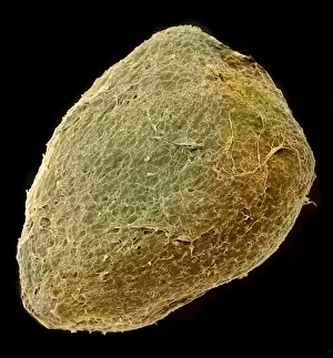
Solanum sp. tomato seedA scanning electron microscope (SEM) image of a tomato seed (Solanum sp.), artificially coloured by computer
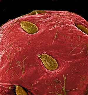
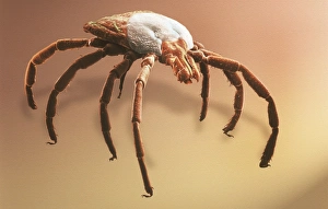
Amblyomma sp. hard backed tickScanning electron microscope view of a hard backed tick from the family Ixodidae. Coloured artificially on computer
Cells on glassScanning electron microscope (SEM) image of cells on glass (x 2K)
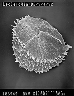
Fractured pollen grainScanning electron microscope (SEM) image showing a fractured pollen grain
Pelargonium crispum, lemon geranium
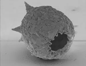
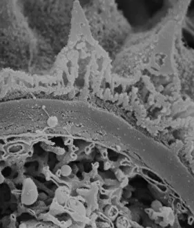
Fractured antherScanning electron microscope (SEM) image showing a fractured anther, otherwise known as the sac, which contains the pollen in the male sex organs (stamens)