mail_outline sales@mediastorehouse.com
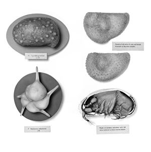
Foraminifera and ostracods modelsBees wax models of foraminifera and ostracods made by Clive Sheppard for an exhibition in the Invertebrates Gallery, at the Natural History Museum, London
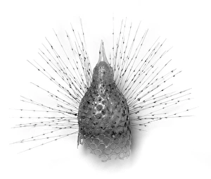
Radiolarian modelGalls model of radiolarian by Blaschka, held at the Natural History Museum, London
Discorbina species, foraminiferaPlate 11 no. 22 of original artwork by Heron-Allen and Earland, 1913, from the Heron-Allen Library at the Natural History Museum, London. Species from the Clare Island Survey, Co. Mayo, Ireland
Foraminifera modelsOne drawer containing some of d Orbigny models and slides previously displayed alongside the models in the galleries
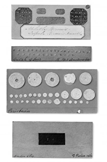
ForaminiferaPart of the display of foraminifera from The Great Exhibition of 1851. Featured are specimens from the London Clay, the Paris Basin and the Gulf of Suez
Difflugia CoronaFreshwater Testate Amoebae. Magnification x 450
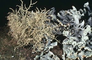
Usnea inflata, beard lichenAnd Hypogymnia physodes (right), Burnham Beeches, Bucks, W. London. The former is a recent colonist following reductions in SO2 pollution
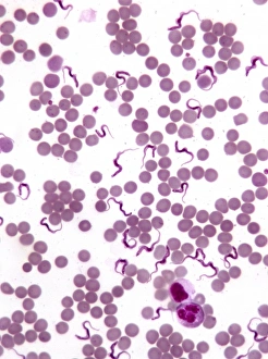
TrypanosomesScanning electron microscope image showing a trypanosoma blood smear. They have proved to be of great interest as they have evolved very differently to other better studied organisms
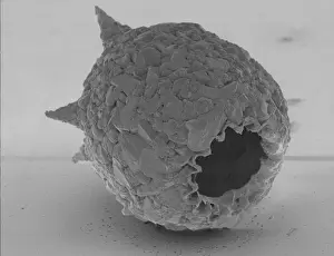
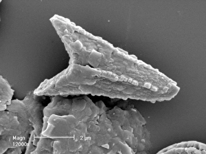
Ceratolithoides aculeus, coccolithScanning electron microscope image of an isolated coocolith from Cretaceous chalk. These are thin calcite shells protecting the coccolithophore within
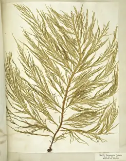
Chorda filum, sea laceCut out of mounted specimen of sea lace or Dead mans rope. A brown seaweed, this specimen is 14.5 feet long and held at the Natural History Museum, London
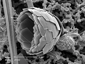
Florosphaera profunda, coccolithScanning electron microscope image of a complete sphere of coccoliths from modern oceans. These are thin calcite shells protecting the coccolithophore within
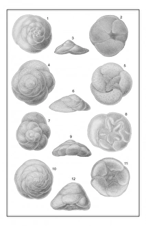
GlobigerinaPlate 77 from Voyage of the H.M.S. Challenger 1873-1876. Zoology Vol. 9. Foraminifera Plates, 1884 by C. Wyville Thomson
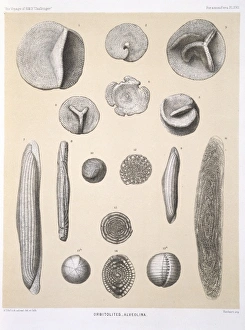
Orbitolites - AlveolinaPlate 17 from Voyage of H.M.S. Challenger (1872-1876). Zoology Vol. 9. Foraminifera Plates, 1884 by C. Wyville Thomson
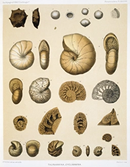
Thurammina - CyclamminaPlate 37 from Voyage of H.M.S. Challenger 1873-1876. Zoology Vol. 9. Foraminifera Plates, 1884 by C. Wyville Thomson
Proterozoic ocean floorA restoration of Proterozoic ocean floor with bun-shaped stromatolites
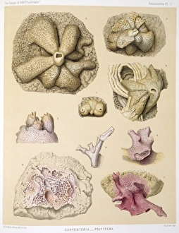
Carpenteria & PolytremaPlate 100 from Voyage of the H.M.S. Challenger 1873-1876. Zoology Vol. 9. Foraminifera Plates, 1884 by C Wyville Thomson
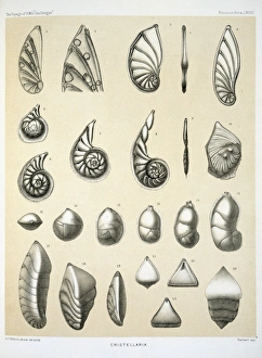
CristellariaPlate 68 from Voyage of the H.M.S. Challenger 1873-1876. Zoology Vol. 9. Foraminifera Plates, 1884 by C. Wyville Thomson
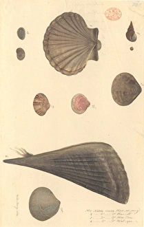
Nine molluscs, including bivalves and gastropodsWatercolour 391 by the Port Jackson Painter, entitled Kow-er-ring, Kow-ill, Kaa-din, Wal-gan, from the Watling Collection
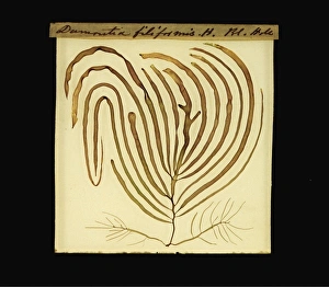
Dumontia contorta, seaweed
Scinaia forcellata, seaweedCut out of specimen of marine alga or seaweed collected by Holmes in Enoura, Japan. Specimen is held in the Crypt. Herbarium at the Natural History Museum, London
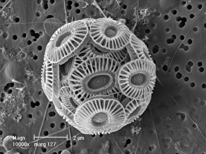
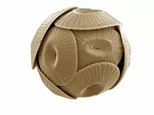
Emiliania huxleyi coccosphereCoccosphere of Emiliania huxleyi from the Western Mediterranean. E. huxleyi is one of the most widespread species on earth
Desmarestia ligulata, seaweedPage 55 from Algae Danmonienses: or dried specimens of Marine Plants, principally collected in Devonshire by Mary Wyatt; carefully named according to Dr. Hookers British Flora
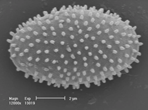
Amphitetras, diatomScanning electron microscope (SEM) image showing the diatom Amphitetras with its ornate silica shell (x5000 on a standard 9 cm wide print). Coloured artificially by computer
Frontispiece of Catalogue Raisonne d une collectionIllustration by Francois Boucher from the book Catalogue Raisonne d une collection by Edme Francois Gersaint, 1744
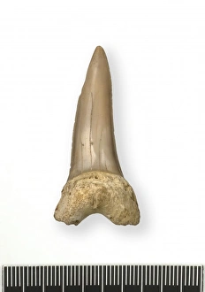
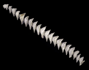
Tonguestone (sharks tooth)A sharks tooth from the species Oxyrhina. Specimen originates from the Globigerina Limestone, Miocene period, NW Malta
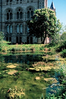
The pond in the Wildlife Garden. Photographed by Derek Adams. Published in Wildlife Garden by Roy Vickery, 2004 page 35
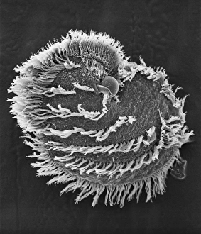
Ciliate planktonScanning electron microscope image of a ciliate showing clearly the microscopic hairs or cilia that they use for movement and feeding (x 700)
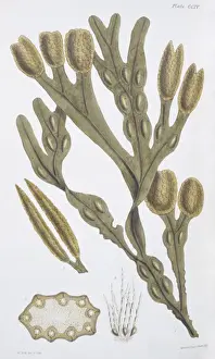
Fucus vesiculosis, bladderwrackIllustration from Botany Library Plate Collection at the Natural History Museum, London. By Leopald Trattinick, 1825
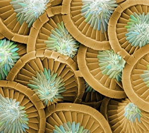
CoccolithsScanning electron microscope (SEM) image of coccoliths, these are the limestone scales surrounding the marine phytoplankton coccolithophores
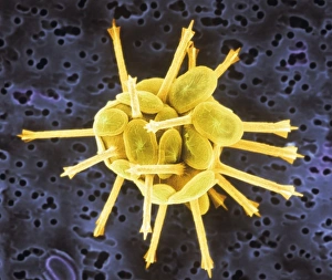
Acanthoica acanthifera
Myxomycetes, plasmodial slime mouldScanning electron microscope image of a plasmodial slime mould spore (x12000). This mould spends most of its life as a single cell; when they reproduce they form a slug-like blob that can travel
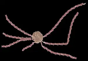
Spiral axis of Archimedes, bryozoanArchimedes, was a bryozoan possibly living in association with an alga. From the Lower Carboniferous limestone, Iowa, USA. c. 350-330 million years old
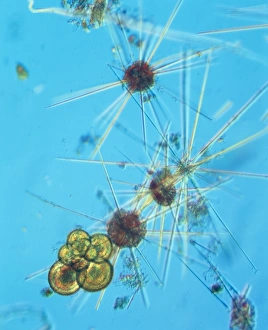
AcanthowetraA photograph of a foraminifera found in the Indian Ocean
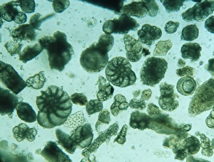
Foraminiferan remains from the White Cliffs of Dover, U.K. The cliffs are made up of unimaginable numbers of chalky shells of long dead marine animals
Coccolithus pelagicusCoccosphere of Coccolithus pelagicus, a common cold water coccolithophore. Collected from the British Continental shelf, North West of Scotland. Specimen diameter 15m. False-coloured SEM image
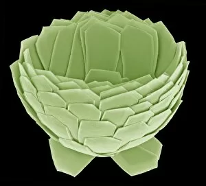
Florisphaera profundaA coccolithophore with highly modified, plate-like coccoliths. This is a very common deep dwelleing species, typically living at about 100-150m depth in the water column
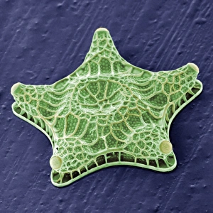
Ophiaster formosusA coccolithophore with long appendages formed of strings of highly modified coccoliths. Collected from the West Pacific. Specimen diameter 50m. False-coloured SEM image