mail_outline sales@mediastorehouse.com
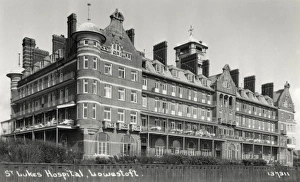
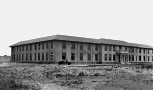
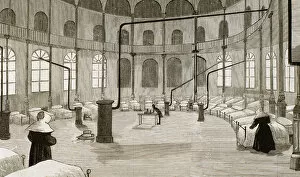
St Lukes Hospital, Lowestoft, SuffolkSt Lukes Hospital at Lowestoft, Suffolk. Formerly the Empire Hotel, the building was acquired in 1921 by the Metropolitan Asylums Board
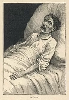
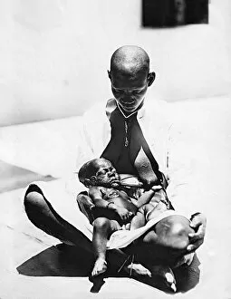
Cholera VictimA cholera victim
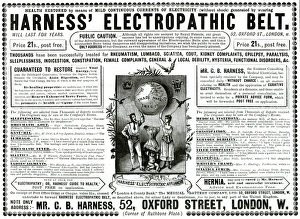
Advert for Harness Electropathic corset belts 1888Harness Electropathic belt treatment. 1888
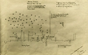
Cholera epidemic, Tiberias, IsraelCholera epidemic, Tiberias, Lower Galilee, Israel, 1-15 October 1918 -- chart drawn by the American Zionist Medical Unit (part of a missionary society), showing cases to the west of Bahar Jehud
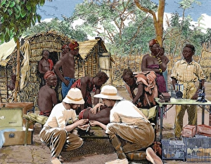
Bacteriology Laboratory, Nairobi, Kenya, East Africa. 1931
Child with Yaws, Mombasa, Kenya, East Africa. 1923
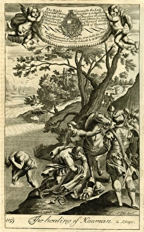
The healing of Naaman - 2 Kings. circa 1690
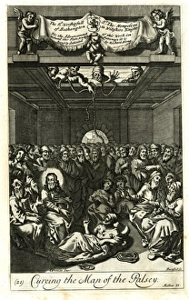
Jesus curing the man of the palsy - Matthew 9. circa 1688
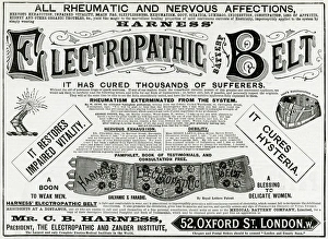
Advert for Harness Electropathic corset belts 1891Advert for Harness Electropathic corset belts no taking poisonous drugs or quack nostrums. Claims to cure all rheumatic and nervous affections. 1891
Spain. Madrid. Womens room on the Hospital installed at theSpain. Madrid. Hospital installed at the Palace of Fine Arts on the occasion of the epidemic of cholera that threatened the city in 1890. Womens room. Engraving
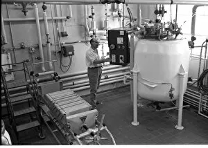
Foot and Mouth Research Laboratory, Pirbright, Surrey, the centre for research into this virulent disease. Seen here is a process worker in the main production unit. Date: 1968
Africa. Native affected by sleeping sickness. Engraving by Thiriat, 1903. Colored
Africa. European doctors examining a native attacked by sleeHISTORY OF AFRICA. Doctors in an expedition of European explorers, examining the native blood attacked by sleeping sickness. Engraving by Thiriat. 1903. Colored
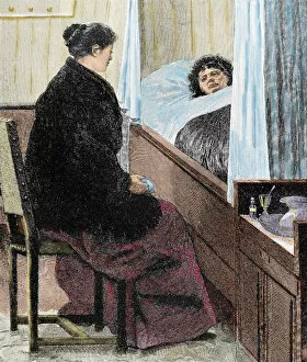
Visiting a sick woman. Colored engraving of The Artistic Illustration, 1892
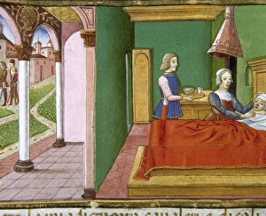
The centurions servant sick in bed. Codex of Predis (1476). Royal Library. Turin. Italy
Abraham Cowley - 3ABRAHAM COWLEY Poet, for whom life is an incurable disease. Date: 1618 - 1667
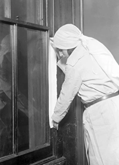
Fumigating a RoomThis young woman covers her mouth to protect herself from breathing in harmful bacteria as she seals a doorway and keyhole of a room in her house for fumigation from disease. Date: early 1930s
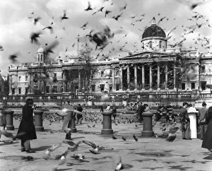
Trafalgar Square BirdsTourists in Trafalgar Square are virtually obscured by the sheer volume of pigeons around them. Date: 1950s
Vernon at Cartagena / 1741The Spanish American port of Cartagena besieged by Admiral Vernon. Despite Vernons claims, the operation was a failure, and many men were lost through disease. Date: March 1741
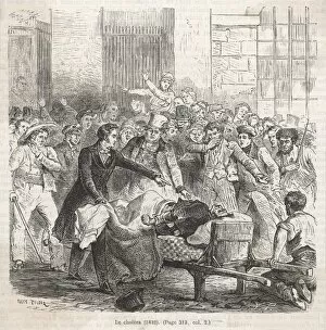
French Cholera victimA horrified crowd gathers round to see a cholera victim in France Date: 1832
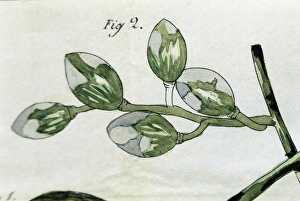
Fragment of a woody branch of vine attacked by the vegetative part of a fungus or mycelium. Illustration for La Maladie des Vignes (The disease of the vines)
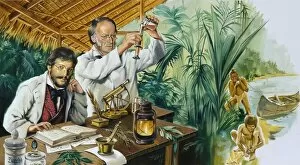
Scientific experiments in the jungle in the early 20th century. Illustration by Lluis Bargall atercolour
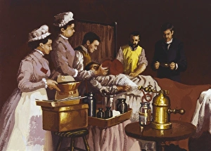
Surgery using ether as anesthetic administered by inhalation, at Bellevue Hospital in New York, 1870. Method first experienced by William T. G. Morton in 1846. Oil
Disease of the vines. Engraving of 1853Bunch of grapes partially attacked by the vegetative part of a fungus or mycelium in the first days of July. Illustration for La Maladie des Vignes (The disease of the vines)
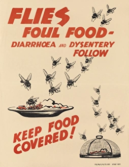
WW2 Poster -- Flies Foul Food -- Keep Food CoveredFlies Foul Food - Diarrhoea and Dysentery Follow. Keep Food Covered! Colour lithograph poster after 2nd Lt Stacey Hopper. Anti-dysentery
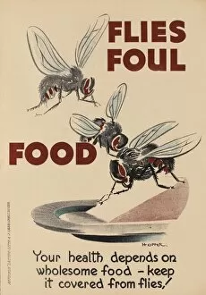
WW2 Poster -- Flies Foul FoodFlies Foul Food. Your health depends on wholesome food - keep it covered from flies! Colour lithograph poster after 2nd Lt Stacey Hopper
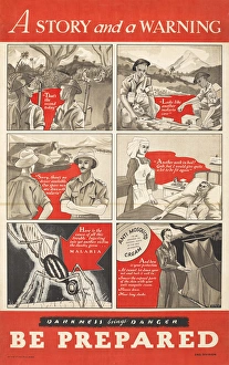
WW2 Poster -- Be PreparedA Story and a Warning, .Darkness brings Danger, Be Prepared. Colour photolithograph poster, published by The Times of India Press, Bombay
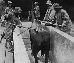
British veterinary hospital in France, 1916Men at a British Veterinary Hospital in France scrubbing a skin disease on a horse after a warm bath during the First World War. Date: 1916
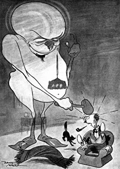
A-Tich-oo! Influenza in 1918Influenza personified in the shape of a rather monstrous being tapping an unsuspecting chap on the head and announcing, Good evening! I m the new influenza. Date: 1918
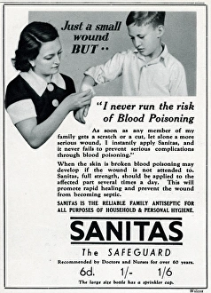
Advert for Sanitas - family antiseptic for wounds 1937Just a small wound but.... I never run the risk of blood poisoning. As soon as any member of my family gets a scratch or cut, let alone a more serious wound, I instantly apply Sanitas
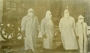
Medical orderlies during plague outbreakMedical orderlies dressed in long coats and gloves and with covered faces, to deal with the bubonic plague outbreak in China around 1911. Date: C.1911
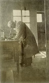
Plague scientist in ChinaWestern doctor or scientist in China during the bubonic plague outbreak; died during research work. Date: 1911
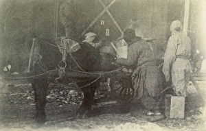
Bubonic plague in ChinaBubonic plague outbreak in China, in Tientsin (now Tianjin): people wrapped up to try to protect themselves from the plague, possibly carrying out some sort of fumigation procedure. Date: 1911
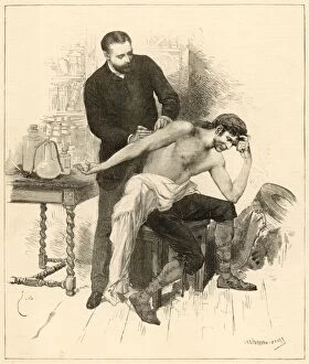
Cholera vaccination in SpainA doctor innoculates a worried gentleman with the cholera vaccine. Date: 1885
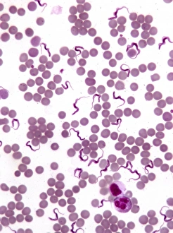
TrypanosomesScanning electron microscope image showing a trypanosoma blood smear. They have proved to be of great interest as they have evolved very differently to other better studied organisms
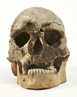
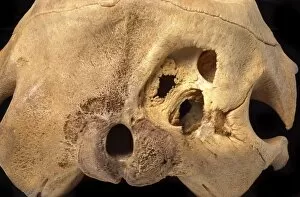
Cheddar ManThe skull of Cheddar Man. This skull is approximately 10, 000 years old. The hole in the forehead may have been caused by infection and also possibly be the cause of death
Ornithodoros moubata, tickThis species of tick (Ornithodoros moubata) specifically carries the virus of African swine fever
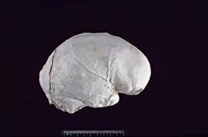
Homo sapiens (Singa 1) cranium endocastAn endocast of a heavily mineralized cranium once belonging to that of Homo sapiens who lived about 130, 000 years ago. This specimen was discovered in Singa, Sudan by W.R.G. Bond in 1924
Kogia breviceps, pygmy sperm whalePhotograph of the skull of a pygmy sperm whale
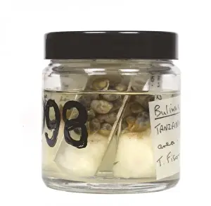
Bulinus sp. snailsSpecimen jar containing snails (Bulinus). These snails act as intermediate hosts for the parasite of the tropical disease bilharzia. Specimens held at the Natural History Museum, London
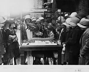
Crowd around flea case, 1927During the first decades of the 20th century, the Central Hall contained a number of exhibit cases explaining the role of insects and other animals in spreading disease
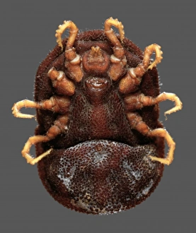
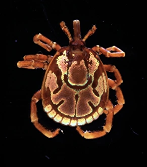
Amblyomma hebraeum, African cattle tickA male African cattle tick (Amblyomma hebraeum). Ticks are blood-sucking parasites wich live off the blood supply from their host
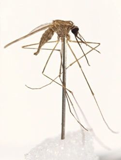
Anopheles labranchiae, mosquitoThis species of mosquito is of medical importance as it is a vector of malaria
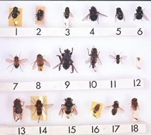
Myiasis speciesA collection of adult Myiasis causing fly specimens. Myaisis is the infestation of organs or tissues of the hosting animals. Photographed by Martin Hall
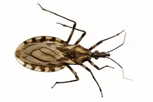
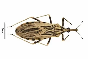
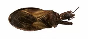
Triatoma brasiliensis, triatomine bugThis insect is a member of the Triatomine group, which are associated with the transmission of disease to humans
Rhodnius ecuadorionsis, triatomine bugThis insect is a member of the Triatomine group, which are associated with the transmission of disease to humans
Cavernicola pilosa, triatomine bugThis insect is a member of the Triatomine group, which are associated with the transmission of disease to humans