mail_outline sales@mediastorehouse.com
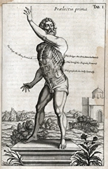
Naked man on plinth with cross section of musclesPraelectio secunda - Tab I. Naked man on plinth with cross section of side and shoulder muscles Source: Myographia nova, or, A graphical description of all the muscles in the humane body
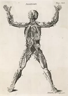
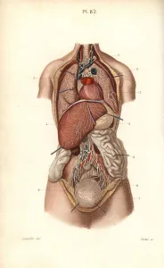
Anatomical drawing of the human bodyAn anatomical drawing of the human body, showing muscles, nerves and internal organs
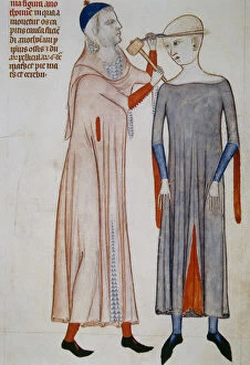
Trepanation. Miniature. 14th c. Treatise on anatomy by MondiIllustration of trepanation. Miniature. 14th century. Treatise on anatomy by Mondino de Liuzzi (1270-1326). Italian anatomist nad profesor of Bologna
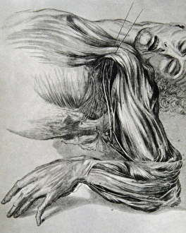
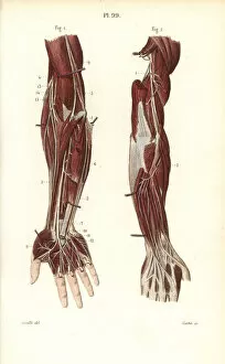
Drawing from a study of anatomy. Human muscles of the armsHistory of medicine. Drawing from a study of anatomy. Human muscles of the arms. 18th century
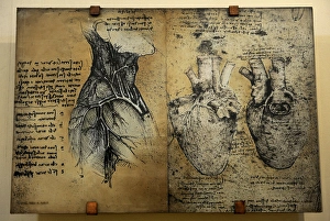
Cardiovascular system Leonardo da vincis drawingCardiovascular systems drawing. In 1513 Leonardo da Vinci to study the heart and the circulatory system through animal dissections. Windsor, Royal Library 19073, 1510-1513
Aquilegia vulgaris, ColumbineInk drawing by Arthur Harry Church, 1903 Date: 1903
Grayling Butterflies Date: 1868
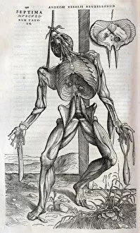
Anatomical drawing of musculatureSeptima Musculorum Tabula (Anatomical drawing of musculature) Source: Andreae Vesalii Bruxellensis, scholae medicorum Patauinae professoris
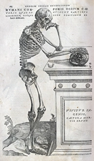
Anatomical drawing of a human skeletonHumani Corporis ossium caeteris quae sustinent partibus liberorum suaque sede positorum (Anatomical drawing of a human skeleton side view) Source: Andreae Vesalii Bruxellensis
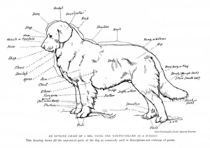
Anatomical diagram of a Newfoundland dog Date: 20th century
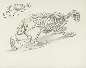
Skeleton of Crouching Lion
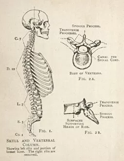
Diagram of human skull and vertebral columnA diagram of the human skull and vertebral column, showing the left ribs and a portion of the breastbone, as well as a single vertebra from two angles
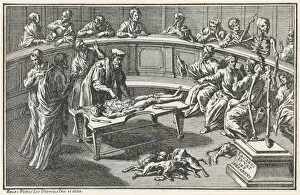
Anatomical Theatre C16Anatomy in the 16th century as depicted in Bartholinus Eustachius works. It looks as though dogs are enjoying the unwanted organs
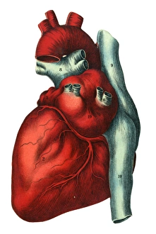
Human heartIllustration of a human heart - top of a layered anatomical fold-out in a medical book. Date: c.1900
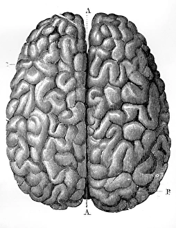
The Human Brain. Date: 1879
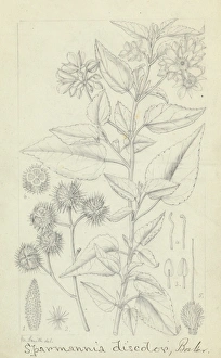
Sparmannia discolor by Matilda SmithGraphite on paper, 1880s by Matilda Smith (1854-1926). Held in the Library and Archives
Deep nervous system to the arm and hand.. Handcolored steel engraving from Dr. Joseph Nicolas Masses Pocket Anatomy of the Human Body, Paris, 1864
Brachial plexus.. Handcolored steel engraving from Dr. Joseph Nicolas Masses Pocket Anatomy of the Human Body, Paris, 1864
Nerves to the arm and hand.. Handcolored steel engraving from Dr. Joseph Nicolas Masses Pocket Anatomy of the Human Body, Paris, 1864
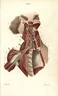
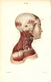
Cervical plexus.. Handcolored steel engraving from Dr. Joseph Nicolas Masses Pocket Anatomy of the Human Body, Paris, 1864
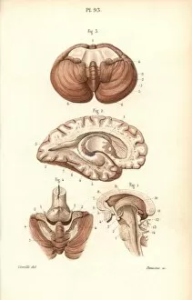
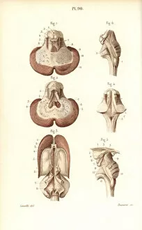
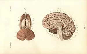
Sections through the brain, cerebellum and ventricles.. Handcolored steel engraving from Dr. Joseph Nicolas Masses Pocket Anatomy of the Human Body, Paris, 1864
Brain, pons, medulla oblongata, pineal gland, etc.. Handcolored steel engraving from Dr. Joseph Nicolas Masses Pocket Anatomy of the Human Body, Paris, 1864
Sections through the brain and pineal gland.. Handcolored steel engraving from Dr. Joseph Nicolas Masses Pocket Anatomy of the Human Body, Paris, 1864
Sections through the brain.. Handcolored steel engraving from Dr. Joseph Nicolas Masses Pocket Anatomy of the Human Body, Paris, 1864
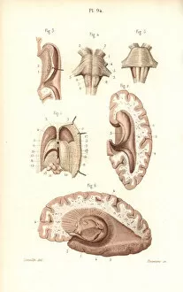
Sections through the cerebellum, ventricular system.. Handcolored steel engraving from Dr. Joseph Nicolas Masses Pocket Anatomy of the Human Body, Paris, 1864
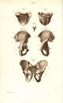
Sacrum, coccyx and pelvis bones.. Handcolored steel engraving from Dr. Joseph Nicolas Masses Pocket Anatomy of the Human Body, Paris, 1864
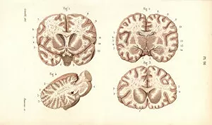
Cross sections through the brain.. Handcolored steel engraving from Dr. Joseph Nicolas Masses Pocket Anatomy of the Human Body, Paris, 1864
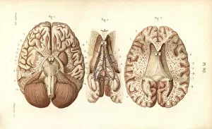
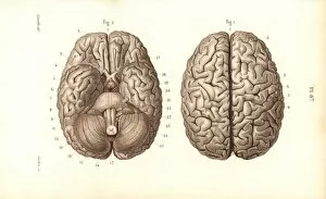
Brain from above and below.. Handcolored steel engraving from Dr. Joseph Nicolas Masses Pocket Anatomy of the Human Body, Paris, 1864
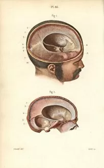
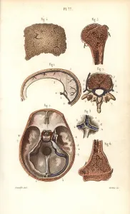
Cross sections through the skull.. Handcolored steel engraving from Dr. Joseph Nicolas Masses Pocket Anatomy of the Human Body, Paris, 1864
Lymph nodes and vessels in the head, neck and chest.. Handcolored steel engraving from Dr. Joseph Nicolas Masses Pocket Anatomy of the Human Body, Paris, 1864
Lymphatic system in the arm.. Handcolored steel engraving from Dr. Joseph Nicolas Masses Pocket Anatomy of the Human Body, Paris, 1864
The thoracic canal.. Handcolored steel engraving from Dr. Joseph Nicolas Masses Pocket Anatomy of the Human Body, Paris, 1864
Lymphatic system to the thorax and abdomen.. Handcolored steel engraving from Dr. Joseph Nicolas Masses Pocket Anatomy of the Human Body, Paris, 1864
Lymph nodes and vessels deep in the back of the leg.. Handcolored steel engraving from Dr. Joseph Nicolas Masses Pocket Anatomy of the Human Body, Paris, 1864
Lymphatic system to the abdomen.. Handcolored steel engraving from Dr. Joseph Nicolas Masses Pocket Anatomy of the Human Body, Paris, 1864
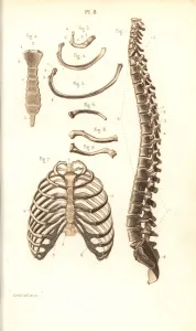
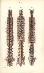
Spine, ribs, vertebrae and sternum.. Handcolored steel engraving from Dr. Joseph Nicolas Masses Pocket Anatomy of the Human Body, Paris, 1864
Lymph nodes and vessels deep in the leg.. Handcolored steel engraving from Dr. Joseph Nicolas Masses Pocket Anatomy of the Human Body, Paris, 1864
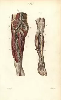
Lymphatic system in the leg and foot.. Handcolored steel engraving from Dr. Joseph Nicolas Masses Pocket Anatomy of the Human Body, Paris, 1864
Sinuses or cavities in skull, humerus and vertebrae.. Handcolored steel engraving from Dr. Joseph Nicolas Masses Pocket Anatomy of the Human Body, Paris, 1864
Veins to the spine.. Handcolored steel engraving from Dr. Joseph Nicolas Masses Pocket Anatomy of the Human Body, Paris, 1864
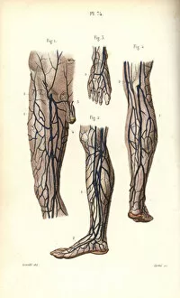
Veins to the leg and foot.. Handcolored steel engraving from Dr. Joseph Nicolas Masses Pocket Anatomy of the Human Body, Paris, 1864
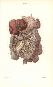
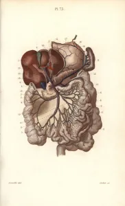
Veins to the stomach, liver and intestines.. Handcolored steel engraving from Dr. Joseph Nicolas Masses Pocket Anatomy of the Human Body, Paris, 1864
Circulatory system to the spine and uterus.. Handcolored steel engraving from Dr. Joseph Nicolas Masses Pocket Anatomy of the Human Body, Paris, 1864
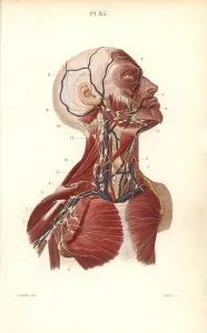
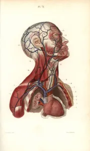
Circulatory system to the head and torso.. Handcolored steel engraving from Dr. Joseph Nicolas Masses Pocket Anatomy of the Human Body, Paris, 1864
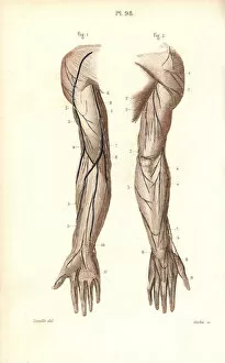
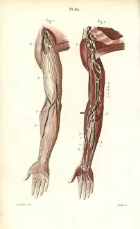
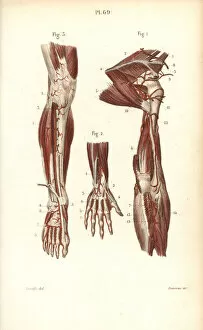
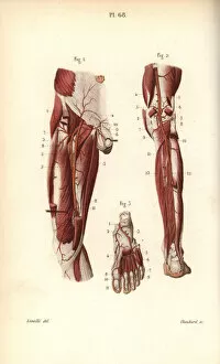
Circulatory system to the arm and hand.. Handcolored steel engraving from Dr. Joseph Nicolas Masses Pocket Anatomy of the Human Body, Paris, 1864
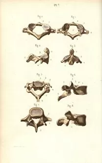
Axis and vertebrae.. Handcolored steel engraving from Dr. Joseph Nicolas Masses Pocket Anatomy of the Human Body, Paris, 1864
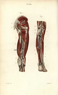
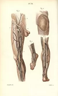
Circulatory system to the leg and foot.. Handcolored steel engraving from Dr. Joseph Nicolas Masses Pocket Anatomy of the Human Body, Paris, 1864
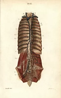
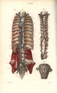
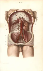
Circulatory system to the abdomen.. Handcolored steel engraving from Dr. Joseph Nicolas Masses Pocket Anatomy of the Human Body, Paris, 1864