mail_outline sales@mediastorehouse.com
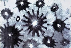
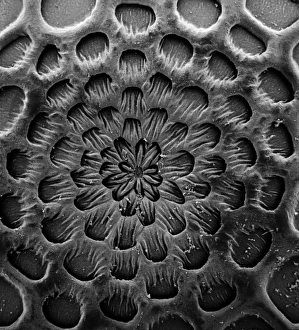
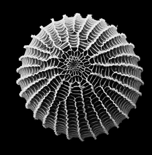
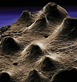
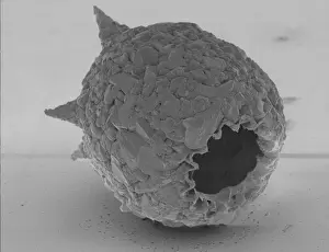
Crysotile asbestosScanning electron micrograph of 5-Fold symmetry in crysotile asbestos. Magnification on the 5 x4 transparency = X 600, 000
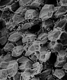
LiverScanning electron microscope (SEM) image of a section through a liver (x 7000), an organ that has over 500 functions in the human body (x 800)
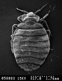
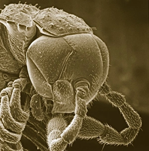
Cimex lectularius, bed bugScanning electron microscope image of a bed bug (x 17). The sucking mouthparts enable the feeding bedbugs to pierce the hosts tissues and siphon out a blood meal
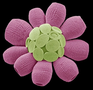
Taraxacum officinale, dandelion (fruiting head)Scanning electron microscope image showing a vertical section through an unripe fruiting head of a dandelion in the yellow flower stage. Colour added artificially by computer
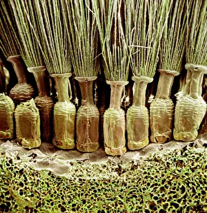
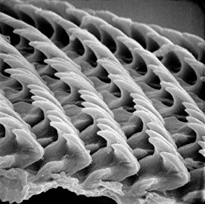
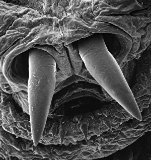
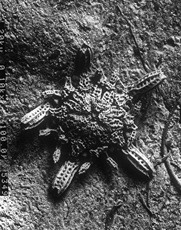
Snail teeth
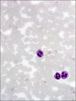
Plasmodium sp. malarial parasiteScanning electron microscope image of a malarial protozoal parasite. The parasite requires the anopheles mosquito to complete its life cycle
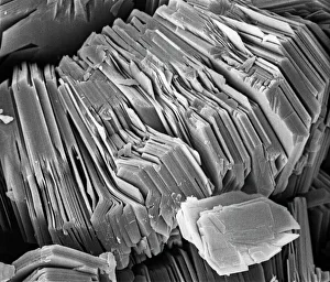
KaoliniteScanning electron microscope image of kaolinite (x 4.00K). Its a common phyllosilicate mineral, its structure is composed of silicate sheets bonded to aluminum oxide/hydroxide layers
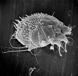
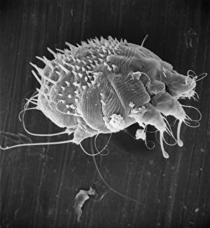
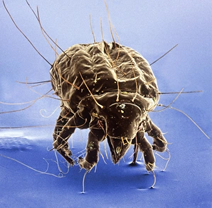
Sarcoptes scabiei, scabies miteScanning electron microscope image of an itch or scabies mite, a parasite that infests a wide variety of mammalian hosts including humans
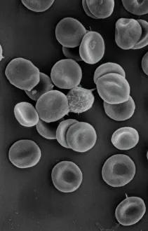
Human red blood corpusclesScanning electron microscope (SEM) of red blood cells showing their characteristic biconcave shape which increases the surface area for diffusion
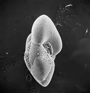
Globorotalia scitula, foraminifera fossilScanning electron microscope (SEM) image showing a fossilised planktonic species of foraminifera
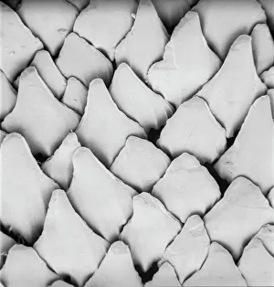
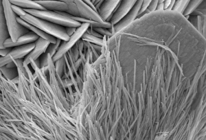
Scyliorhinus canicula, dogfishScanning electron microscope (SEM) image of the scales of a dogfish (x 40)
AspergillusAn SEM image of aspergillus in spore production (x 815 on a standard 9 cm wide print). The moulds are common in the northern hemisphere and some cause disease in humans and animals
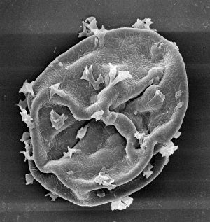
Caterpillar eggScanning electron microscope image of a caterpillar egg (x 90), the caterpillar emerges by chewing through the shell (x 350)
Blackfly antennaScanning electron microscope image of a blackfly antenna (x 350). These long sensory organs feel and taste objects as well as sensing vibrations and smells (x 1.1K)
Anopheles gambiae, mosquitoScanning electron microscope image showing a close-up of the compound eye of a female mosquito (x 2200 on a standard 9 cm wide print)
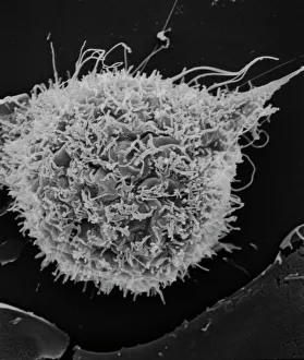
T2 cell cultureScanning electron microscope image showing a T2 cell culture (x 4K)
The anus of a bot flyScanning electron microscope image of the anus of a bot fly. Image on display in the Darwin Centre at the Natural History Museum, London
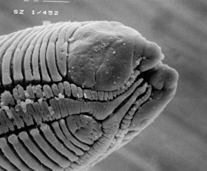
RoundwormScanning electron microscope (SEM) image of a parasitic roundworms head (x 1000 on a standard 9 cm wide print)
Moth eggScanning electron microscope (SEM) image of a moth egg (x 90). The caterpillar emerges by chewing through the shell
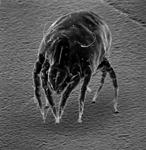
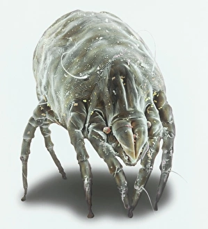
Dermatophagoides sp. dust miteScanning electron microscope image of a dust mite. Dust mites are secondary to pollen as a cause for allergies and they live in bedding, soft furniture and carpets
Sarcoptes scabiei, scabies miteScanning electron microscope image of an itch or scabies mite, a parasite that infests a wide variety of mammalian hosts including man
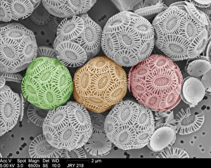
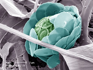
Emiliania huxleyi coccolithophores collected from a bloom in the SW Approaches to the English Channel in June 2004. Date: 2004
Sea saltA scanning electron microscope (SEM) image of sea salt, artificially coloured by computer
Tyrophagus casei, cheese miteScanning electron microscope image of a cheese mite (x 170). These creatures are generally considered to be a pest, however they are added to Altenburger cheese to give it flavour
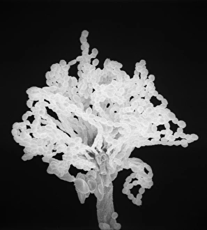
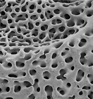
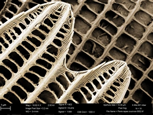
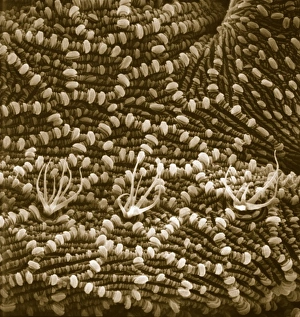
SEM of echinoderm steroemA SEM of an example of stereom of an echinoderm (phylum which consists of 5 classes including starfish). Stereom is the structure formed by the fine networks of calcium carbonate which constitute
Dermanyssus gallinae, red or poultry miteScanning electron microscope image of the red or poutry mite. Adults appear red when engorged with blood, but otherwise are black, grey or white. Females are about 1mm long
Ceratodon purpureus, ceratodon moss spore capsuleScanning electron microscope (SEM) image of a ceratodon moss spore capsule (x 650 on a standard 9 cm wide print)
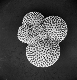
ForaminiferScanning electron microscope (SEM) image of a foraminifer - a single celled organism
Dermatophagoides pteronyssius, dust miteScanning electron microscope image showing a dust mite (x 250 on standard 9cm wide print). This image has been artificially coloured by a computer
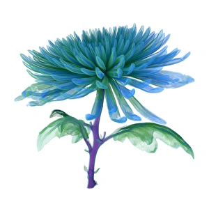
Chrysanthemum, CT scan imageCT Scan image of a Chrysanthemum
Syracosphaera anthosCoccosphere from the Western Mediterranean. False coloured to show the shell is formed of inner and outer layers of coccoliths with very different structure
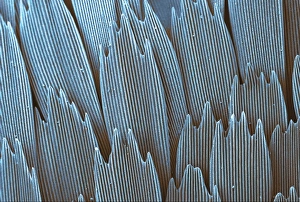
Papilio palinurus, emerald swallowtailSEM image of an emerald swallowtails wing
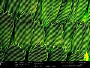
Pieris rapae, small whiteSEM image of the wing of a small white butterfly
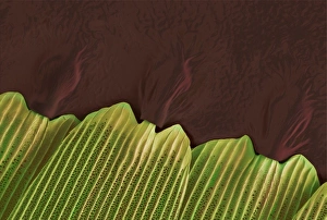
Papilio machaon, old world swallowtailSEM image of Papilio machaon wing
Malachite comprises of (copper carbonate hydroxide). Malachite has distinctive green banding and belongs to the carbonate class
Scyphosphaera apsteinii. SEM image of an equatorial coccolith
Dinosaur eggshellScanning electron microscope image on display in the Darwin Centre
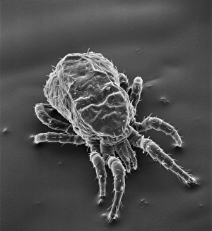
Ventral surface of a mite from the prostigmatic speciesScanning electron microscope image displayed on the glass screens in the Darwin Centre, at the Natural History Museum, London
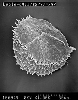
Visbyshaera oligofurcata, acritarchScanning electron microscope image of a microfossil belonging to a group of marine phytoplanktonic organisms known as acritarchs that teemed in Silurian seas about 415 Ma ago
Oestridae, botfly larvaScanning electron microscope image of a botfly larva. They are parasites feeding on skin in the case of warble flies, nostrils in the flies that affect sheep and deer
Difflugia CoronaFreshwater Testate Amoebae. Magnification x 450
Lycopod
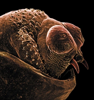
Diplopoda sp. plate millipedeScanning electron microscope image of a lateral view of the head of a plate millipede. Image displayed on the glass screens in the Darwin Centre, at the Natural History Museum, London
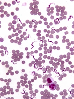
TrypanosomesScanning electron microscope image showing a trypanosoma blood smear. They have proved to be of great interest as they have evolved very differently to other better studied organisms
Ophioctenella sp. brittle starScanning electron microscope image of the post-larval stage of a brittle star (x 110) A newly described species 1994