mail_outline sales@mediastorehouse.com
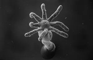
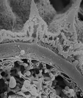
Hydra spScanning electron microscope (SEM) image showing the stinging tentacles and mouth of the coelenterate Hydra (x 36 on a standard 9cm wide print)
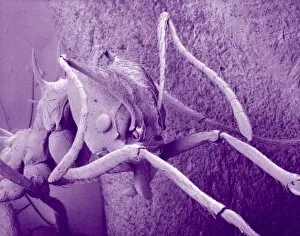
Atta cethalotes, leaf-cutter antScanning electron microscope image of a leaf-cutter ant displayed in the Darwin Centre, at the Natural History Museum, London
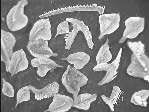
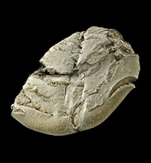
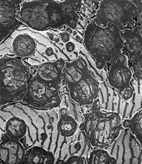
Conodont fossilsScanning electron microscope image of fossils from the Devonian period of northern Estonia, about 465 Ma old ( x 4.2). These creatures are still a mystery to paleontologists
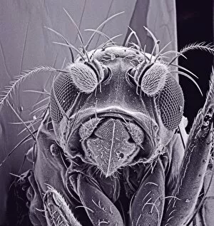
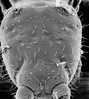
Small fly, species unknownScanning electron microscope (SEM) of a fly head. Image displayed on the glass screens in the Darwin Centre, at the Natural History Museum, London
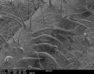
Pelargonium sp. geraniumScanning Electron Microscope image of a pelaronium leaf
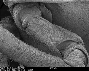
Woodlouse antennaScanning Electron Microscope (SEM) image of woodlouse antenna
OatsA scanning electron microscope (SEM) image of oats, artificially coloured by computer
Spinacia oleracea, spinachA scanning electron microscope (SEM) image of spianch (Spinacia oleracea), artificially coloured by computer
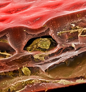
Solanum sp. tomatoA scanning electron microscope (SEM) image of a tomato (Solanum sp.), artificially coloured by computer
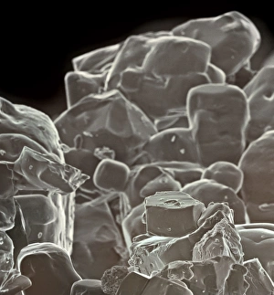
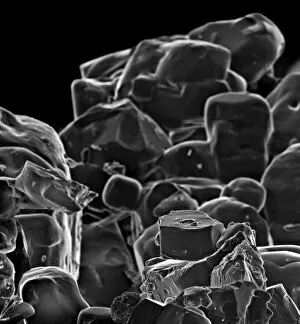
Table saltA scanning electron microscope (SEM) image of table salt, artificially coloured by computer
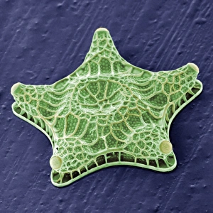
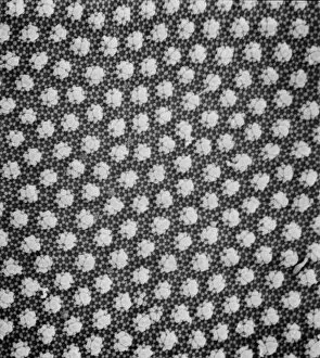
Amphitetras, diatomScanning electron microscope (SEM) image showing the diatom Amphitetras with its ornate silica shell (x5000 on a standard 9 cm wide print). Coloured artificially by computer
Vitis sp. red grapeA scanning electron microscope (SEM) image of a red grape (Vitis sp.), artificially coloured by computer
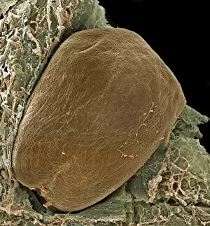
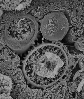
Vitis sp. grape seedScanning electron microscope (SEM) image of a grape seed (Vitis sp.), artificially coloured by computer
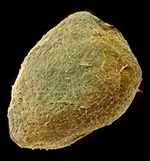
Solanum sp. tomato seedA scanning electron microscope (SEM) image of a tomato seed (Solanum sp.), artificially coloured by computer
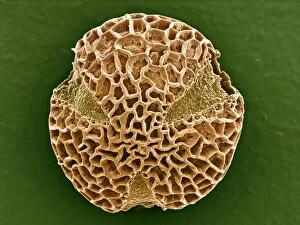
Leptoglossis ferreyraeiA pollen grain of Leptoglossis ferreyraei (polar view) from the family Solanaceae, the tomato family
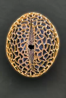
Leptoglossis lomanaA pollen grain of the Leptoglossis lomana (polar view) from the family Solanacea, the tomato family
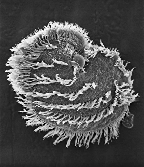
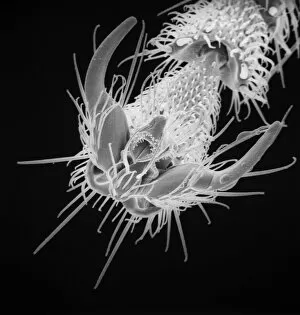
Ciliate planktonScanning electron microscope image of a ciliate showing clearly the microscopic hairs or cilia that they use for movement and feeding (x 700)
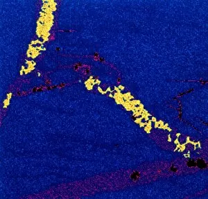
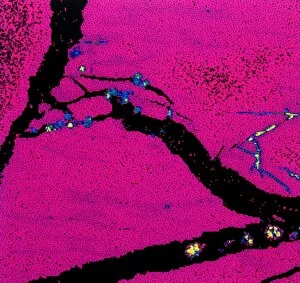
Gold in unspecified mineralScanning electron microscope image of an elemental map showing the distribution of gold (Au) in mineral samples
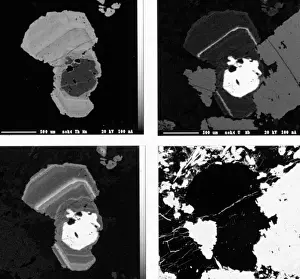
PyrochloreScanning electron microscope images of elemental maps showing thorium, uranium, tantalum and silicon in the mineral pyrochlore from Sokli, Finland
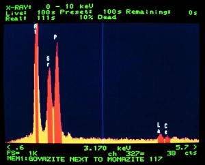
GoyaziteScanning electron microscope image of the energy-dispersive X-ray spectrum of the mineral goyazite, obtained using Link AN10000 analysis system
Collembola, springtailScanning electron microscope image of a springtail head (x 300)
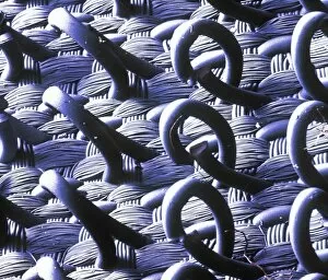
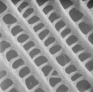
VelcroA trademarked name for a fastening tape made up of a strip of nylon with a surface of minute hooks, that fasten to another strip with a surface of uncut pile. A SEM image
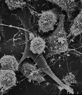
Cells on glassScanning electron microscope (SEM) image of cells on glass (x 2K)
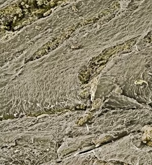
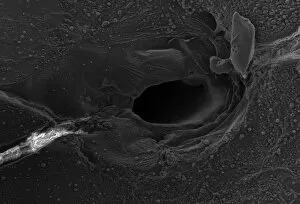
Volcanic glass, Peles hairScanning electron microscope image of a sample of volcanic glass from Mt. Pele, produced to evaluate different types of laser in Laser Ablation Inductively Coupled Plasma Mass Spectrometry
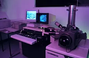
Variable pressure scanning electron microscopeThis electron microscope allows the imaging of samples without any preparation
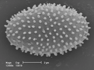
Myxomycetes, plasmodial slime mouldScanning electron microscope image of a plasmodial slime mould spore (x12000). This mould spends most of its life as a single cell; when they reproduce they form a slug-like blob that can travel
Collembola ocelli, springtailScanning electron microscope image of the springtail with simple eyes (x 1.2K)
Copper in unspecified mineralScanning electron microscope image of an elemental map showing the distribution of copper (Cu) in mineral samples
OstracodScanning electron microscope image of an ostracod, an arthropod where the body is enclosed in a carapace (x 220)
Collembola sp. springtailScanning electron microscope image of a springtail showing the characteristic pattern on the cuticle surface (x 3.5K)
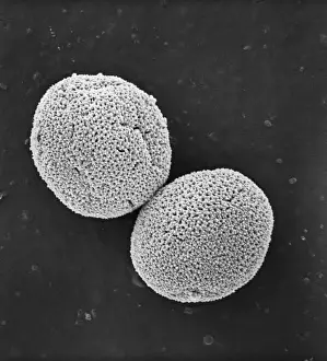
Fractured pollen grainScanning electron microscope (SEM) image showing a fractured pollen grain
Populus nigra, lombardy or black poplar pollenScanning electron microscope image (x 1500) of black poplar pollen grains showing a characteristic granular surface ornamentation and no apertures (inaperturate)
Butterfly wing scale (part)
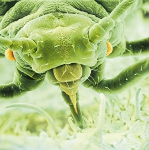
Aphis fabae, black bean aphidScanning electron microscope image showing a frontal view of a black bean aphid on leaf (x100). Aphids or plant lice are small, plant-sucking insects
Human hairScanning electron microscope (SEM) image showing a human hair with the cuticle reflexed
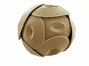
Coccolithus pelagicusCoccosphere of Coccolithus pelagicus, a common cold water coccolithophore. Collected from the British Continental shelf, North West of Scotland. Specimen diameter 15m. False-coloured SEM image
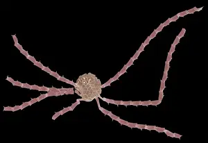
Ophiaster formosusA coccolithophore with long appendages formed of strings of highly modified coccoliths. Collected from the West Pacific. Specimen diameter 50m. False-coloured SEM image
Lasius niger, black garden ant
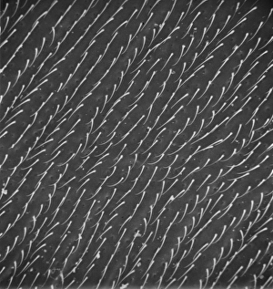
Calliphora vicina, blowfly or blue bottleScanning electron microscope (SEM) image of a blowflys wing
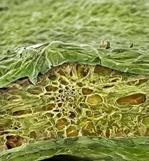
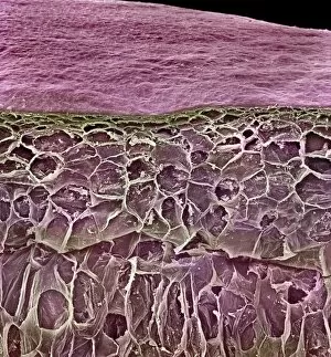
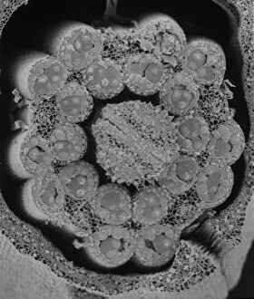
Fractured antherScanning electron microscope (SEM) image showing a fractured anther, otherwise known as the sac, which contains the pollen in the male sex organs (stamens)
Asteraceae, daisyScanning electron microscope image of the fractured surface of an anther showing a developing pollen grain from a member of the daisy or Asteraceae family ( X 3000)
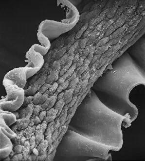
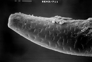
Lumbricus terrestris, earthwormScanning electron microscope (SEM) image showing the chaeta/setae - involved in the locomotion on an earthworm