mail_outline sales@mediastorehouse.com
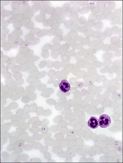
Plasmodium sp. malarial parasiteScanning electron microscope image of a malarial protozoal parasite. The parasite requires the anopheles mosquito to complete its life cycle
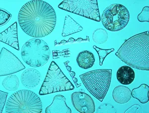
DiatomsSelected slide of a group of fossil diatoms collected from Bori, Hungary in September 1895 and viewed under the light microscipe using differential interfereance contrast
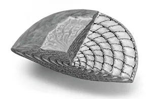
Foraminifer modelModel of typical nummulitic foraminfer after Zittel
Amoeba proteus, amoebaeA glass model of amoebae, created by Leopold and Rudolf Blaschka in the late nineteenth century and held at the Natural History Museum, London
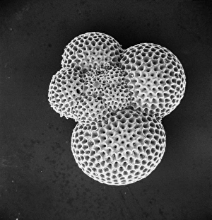
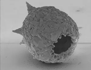
ForaminiferScanning electron microscope (SEM) image of a foraminifer - a single celled organism
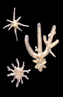
Dorataspis diodon, radiolarianA glass model of a radiolarian, created by Leopold and Rudolf Blaschka in the late nineteenth century and held at the Natural History Museum, London
Aulacantha scolymantha, radiolarianA glass model of a radiolarian, created by Leopold and Rudolf Blaschka in the late nineteenth century and held at the Natural History Museum, London
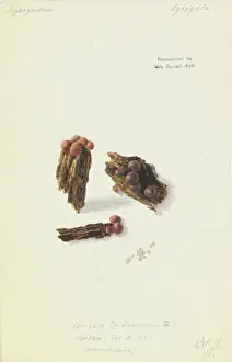
Lycogala epidendrum, Wolfs MilkWatercolour on paper, c.1838 by Anna Russell (nee Worsley) (1807-1876). Held in the Library and Archives Date: circa 1838
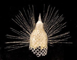
Eucyrtidium cranoides, radiolarianA glass model of a radiolarian, created by Leopold and Rudolf Blaschka in the late nineteenth century and held at the Natural History Museum, London
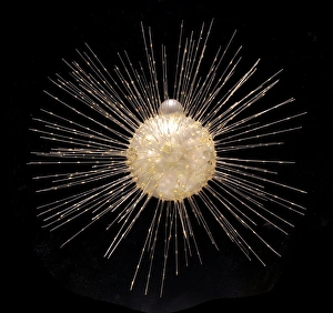
Actinophrys sol, heliozoanA glass model of a radiolarian, created by Leopold and Rudolf Blaschka in the late nineteenth century and held at the Natural History Museum, London
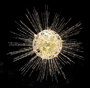
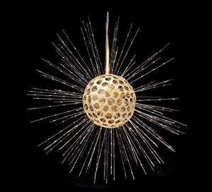
Heliosphaera actinota, radiolarianA glass model of a radiolarian, created by Leopold and Rudolf Blaschka in the late nineteenth century and held at the Natural History Museum, London
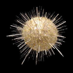
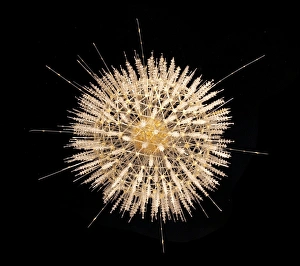
Aulosphaera elegantissima, radiolarianA glass model of a radiolarian, created by Leopold and Rudolf Blaschka in the late nineteenth century and held at the Natural History Museum, London
Difflugia pyriformis, amoebaeA glass model of amoebae, created by Leopold and Rudolf Blaschka in the late nineteenth century and held at the Natural History Museum, London
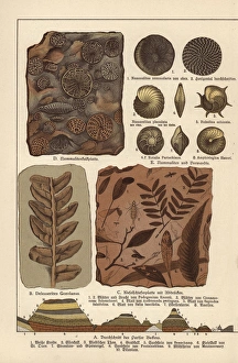
Fossils of algae, plants, insects and protozoa.. Chromolithograph from Dr. Fr. Rolles Geology and Paleontology section in Gotthilf Heinrich von Schuberts Natural History, Schreiber, Munich, 1886
Vorticella convallaria, protozoan.. Handcolored copperplate engraving from George Shaw and Frederick Nodders The Naturalists Miscellany, London, 1797
Foraminifera and ostracods modelsBees wax models of foraminifera and ostracods made by Clive Sheppard for an exhibition in the Invertebrates Gallery, at the Natural History Museum, London
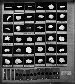
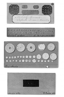
Foraminifera modelsOne drawer containing some of d Orbigny models and slides previously displayed alongside the models in the galleries
ForaminiferaPart of the display of foraminifera from The Great Exhibition of 1851. Featured are specimens from the London Clay, the Paris Basin and the Gulf of Suez
Difflugia CoronaFreshwater Testate Amoebae. Magnification x 450
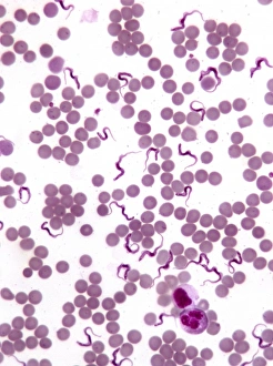
TrypanosomesScanning electron microscope image showing a trypanosoma blood smear. They have proved to be of great interest as they have evolved very differently to other better studied organisms
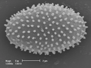
Myxomycetes, plasmodial slime mouldScanning electron microscope image of a plasmodial slime mould spore (x12000). This mould spends most of its life as a single cell; when they reproduce they form a slug-like blob that can travel
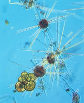
AcanthowetraA photograph of a foraminifera found in the Indian Ocean
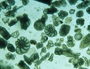
Foraminiferan remains from the White Cliffs of Dover, U.K. The cliffs are made up of unimaginable numbers of chalky shells of long dead marine animals
Minakatella longifila, slime mould