mail_outline sales@mediastorehouse.com
Cystopteris diaphana, diaphanous bladder fernAn SEM showing a close-up of the spiny-lacunar surface of the diaphanous bladder fern (Cystopteris diaphana) spore. Photographed using Philips XL30 SEM
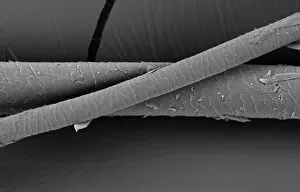
Hair of the DogA scanning electron micrograph (SEM) of a dog hair
Mustelus mustelus, smoothhound sharkA Scanning Electron Microscope image of smoothhound shark skin. The skin is covered with tiny teeth called dermal denticles
Fragaria sp. strawberryA scanning electron microscope (SEM) image of a strawberry (Fragaria sp.), artificially coloured by computer
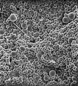
Sugar grainsA scanning electron microscope (SEM) image of sugar grains, artificially coloured by computer
Vitis sp. white grapeA scanning electron microscope (SEM) image of a white grape (Vitis sp.), artificially coloured by computer
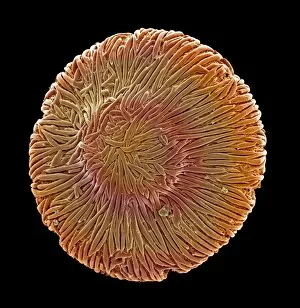
Browallia speciosa, amethystA pollen grain of the Browallia speciosa (polar view) from the family Solanaceae, the tomato family
Vaccinium sp. blueberryA scanning electron microscope (SEM) image of a blueberry (Vaccinium sp.), artificially coloured by computer
Solanum sp. tomatoA scanning electron microscope (SEM) image of a tomato (Solanum sp.), artificially coloured by computer
Isurus oxyrinchus, mako sharkScanning Electron Microscope image of mako shark skin
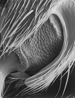
Lasius niger, black garden antScanning electron microscope (SEM) of a black ant leg. Widespread and common in a range of habitats but perhaps most familiar in gardens where nests are formed under paving stones and brickwork
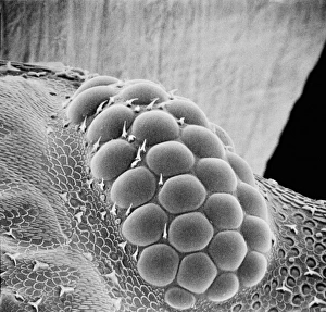
Porcellio sccaber, woodlouseScanning electron microscope (SEM) image showing all the units that make up the compound eye of a woodlouse
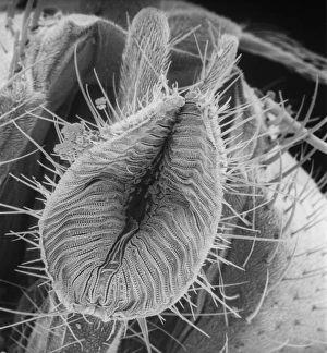
Calliphora vicina, blowfly or bluebottleScanning electron microscope (SEM) image of a blowfly proboscis (x 85). This specialised mouth-part is used to squirt digestive enzymes onto the food
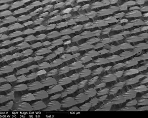
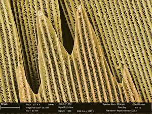
Papilio machaon, old world swallowtailSEM image of a Papilio machaon wing
Microscope image of the Johnstown diogenite. Diogenites are coarse grained and composed primarily of one mineral, pyroxene. Field of view is 2.5mm across
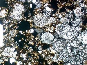
Microscope image of chondrite showing chondrules, typical of primitive meteorites. Horizontal field of view, 3.3mm
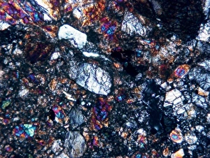
Microscope image of the Zagami shergottite. The fractures in the pyroxene mineral grains and the paler patches of glass show that the rock has been shocked. Field of view is 5mm
Microscope image of the Brachina meteorite, the type specimen of the Brachinite meteorites. Brachinites are composed mostly of olivine with minor amounts of pyroxene and plagioclase
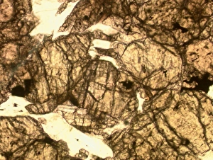
Microscope image of the Lodran meteorite. This meteorite is the type specimen of the Lodranite meteorites. The lodranites are related to the acaplucoites but are more course-grained
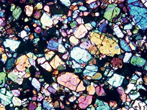
Optical microscope image of the Barwell (Type 6) chondrite. This meteorite has experienced a significant amount of heating
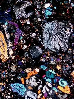
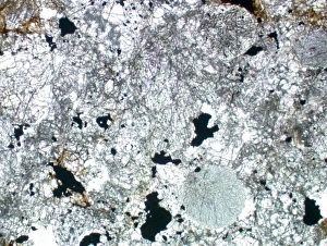
Optical microscope image of the Parnallee (Type 3) chondriteAn optical microscope image of the Parnallee (Type 3) chondrite that has experienced little heating. The chondrules are clear and well-defined. The field of view is 5mm
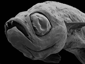
Danionella dracula, minnowSEM image of the Danionella dracula. This tiny 17mm fish has evolved many unique and unusual characteristics, the most spectacular of which are jaw modifications that resemble true teeth
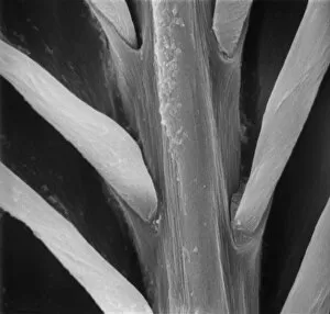
Feather detail
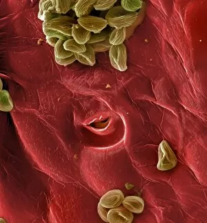
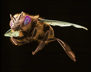
Pollen on beeScanning electron microscope (SEM) image of pollen on a bee. If the plant depends on animals for pollination, the pollen will be relatively large and sticky
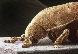
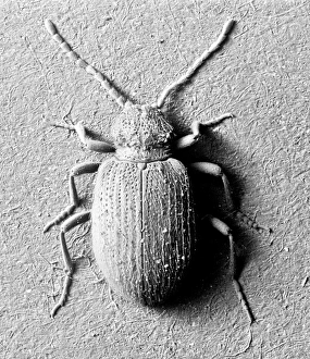
Dermestes lardarius, larder beetleScanning electron microscope image of a larder beetle (x22). These beetles are important for the damage they do, mainly through feeding on animal matter. Coloured artificially by computer
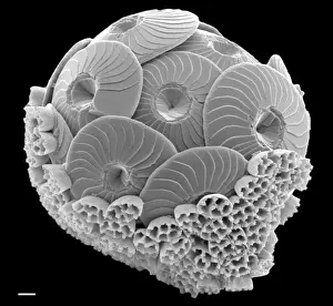
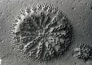
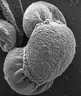
Calcidiscus leptoporus and Syracolithus quadriperforatus, coIn this scanning electron micrograph, the transition of a life-cycle stage in Calcidiscus is shown from the outer cover to the inner layer. Specimen taken from W. Mediterranean
Apis mellifera, honey beeScanning electron microscope image of a honey bee coloured artificially by computer. The female worker caste of this species have special baskets on their legs to to take pollen back to the nest
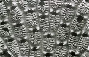
Actinopora disticha, bryozoanScanning electron micrograph of a fossil cyclostome bryozoan from the Cretaceous Chalk, Santonian, Kent
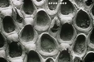
Wilbertopora woodwardi (Brydone), bryozoanScanning electron micrograph of a fossil cheilostome bryozoan. Specimen originates from the Upper Cretaceous Chalk, West Mean Station, Hampshire, U.K
Ptinus tectus, spider beetleScanning electron microscope image of a spider beetle (x 9). The long antennae, hairy body and waist-like constriction give this beetle the appearance of a spider
Aspidelectra melolontha, bryozoanScanning electron micrograph. Zooids of a bleached colony of a modern cheilostome bryozoan. A recent specimen from Sheppey, Kent
Pinus sylvestris, scots pineScanning electron microscope (SEM) image showing a pollen grain from a scots pine. Note the air bladders that help it to float through the air (x 1500 on a standard 9 cm wide print)
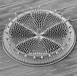
Actinoptychus, diatomScanning electron microscope image of the exterior valve of the diatom Actinoptychus (x 500 on a standard 9 cm wide print)
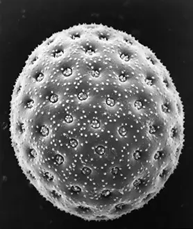
Chenopodium album, goosefootScanning electron microscope image of a pollen grain from a member of the goosefoot family (x 3000 on a standard 9 cm wide print)
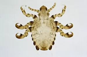
Pthirus gorillae, gorilla lousePhotomicrograph of a gorilla louse specimen, length 2.5 mm from Rwanda / Zaire. The gorilla louse is from the same genus as the human louse
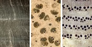
Gymnosperm, palm & angiospermComposite photomicrograph comparing structures visible through a hand-held lens in transverse section of fossil tree trunks: gymnosperm (left), palm (centre) and angiosperm (right)
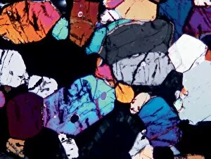
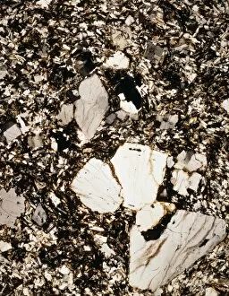
Granite from Ailsa CraigA photomicrograph of granite taken between crossed polarisers. Granite is an igneous rock