mail_outline sales@mediastorehouse.com
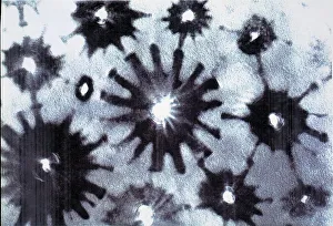
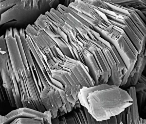
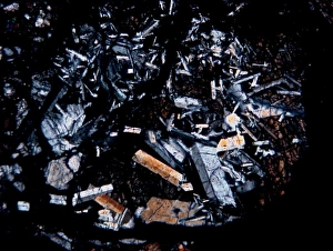
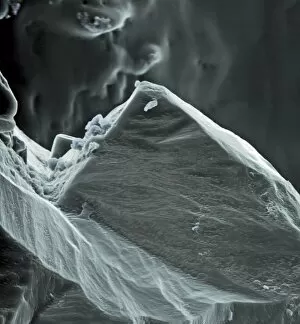
Crysotile asbestosScanning electron micrograph of 5-Fold symmetry in crysotile asbestos. Magnification on the 5 x4 transparency = X 600, 000
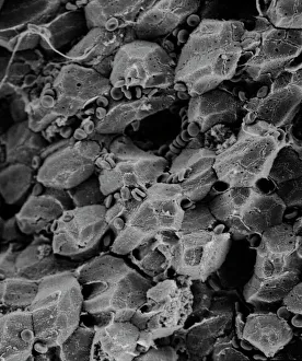
LiverScanning electron microscope (SEM) image of a section through a liver (x 7000), an organ that has over 500 functions in the human body (x 800)
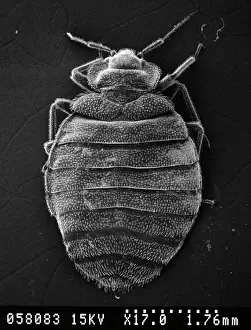
Cimex lectularius, bed bugScanning electron microscope image of a bed bug (x 17). The sucking mouthparts enable the feeding bedbugs to pierce the hosts tissues and siphon out a blood meal
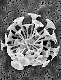
Discosphaera tubifera, coccolithophoreScanning electron microscope (SEM) showing the unicellular planktonic algae Discosphaera tubifera from the North Atlantic surrounded by a sphere of calcite plates - coccoliths
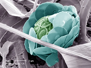
Taraxacum officinale, dandelion (fruiting head)Scanning electron microscope image showing a vertical section through an unripe fruiting head of a dandelion in the yellow flower stage. Colour added artificially by computer
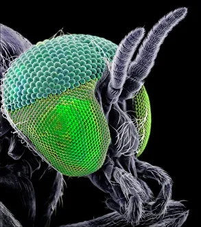
Simulium damnosum, Simulian blackflyScanning electron microscope image of the head showing the compound eye (x 130). The fly is a vector of a parasite which causes River Blindness. Coloured artifically by computer
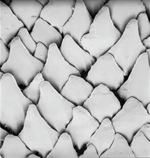
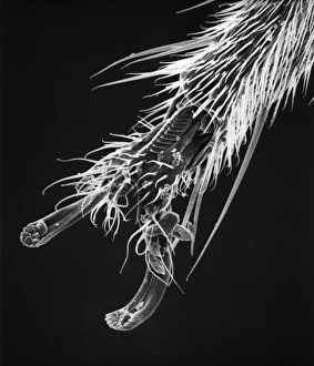
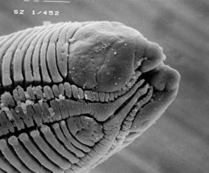
Snail teeth
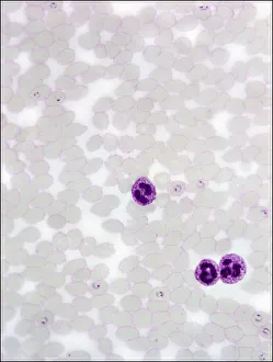
Plasmodium sp. malarial parasiteScanning electron microscope image of a malarial protozoal parasite. The parasite requires the anopheles mosquito to complete its life cycle
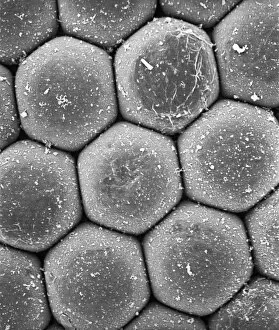
KaoliniteScanning electron microscope image of kaolinite (x 4.00K). Its a common phyllosilicate mineral, its structure is composed of silicate sheets bonded to aluminum oxide/hydroxide layers
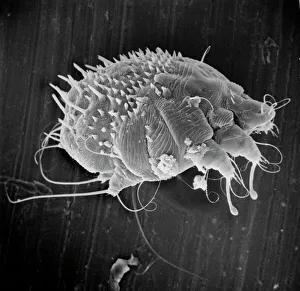
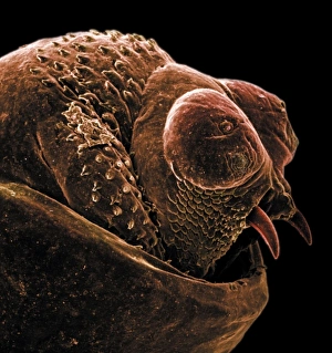
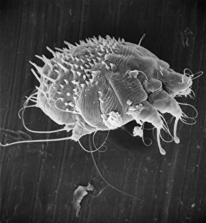
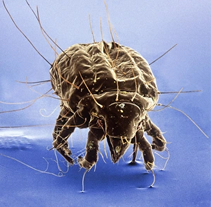
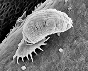
Sarcoptes scabiei, scabies miteScanning electron microscope image of an itch or scabies mite, a parasite that infests a wide variety of mammalian hosts including humans
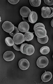
Human red blood corpusclesScanning electron microscope (SEM) of red blood cells showing their characteristic biconcave shape which increases the surface area for diffusion
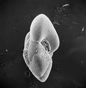
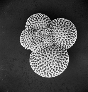
Globorotalia scitula, foraminifera fossilScanning electron microscope (SEM) image showing a fossilised planktonic species of foraminifera
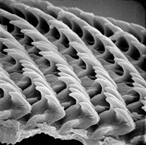
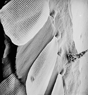
Scyliorhinus canicula, dogfishScanning electron microscope (SEM) image of the scales of a dogfish (x 40)
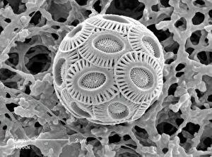
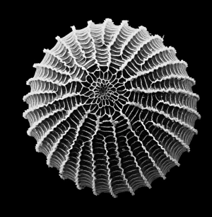
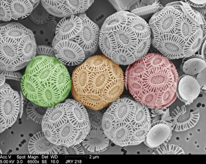
Emiliana huxleyi, coccolithScanning electron microscope image of a complete sphere of coccoliths from modern oceans. These are thin calcite shells protecting the coccolithophore within
AspergillusAn SEM image of aspergillus in spore production (x 815 on a standard 9 cm wide print). The moulds are common in the northern hemisphere and some cause disease in humans and animals
Caterpillar eggScanning electron microscope image of a caterpillar egg (x 90), the caterpillar emerges by chewing through the shell (x 350)
Blackfly antennaScanning electron microscope image of a blackfly antenna (x 350). These long sensory organs feel and taste objects as well as sensing vibrations and smells (x 1.1K)
Anopheles gambiae, mosquitoScanning electron microscope image showing a close-up of the compound eye of a female mosquito (x 2200 on a standard 9 cm wide print)
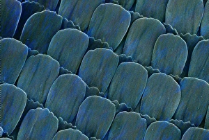
Morpho menelaus, blue morphoScanning electron microscope image of the wing scales from the wing of a South American blue morpho butterfly (x 670 on a standard 9 cm wide print)
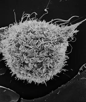
T2 cell cultureScanning electron microscope image showing a T2 cell culture (x 4K)
Microscope image of the Pasamonte eucriteMicroscopic image of the Pasamonte eucrite showing a basaltic texture. Field of view is 2.5mm across
The anus of a bot flyScanning electron microscope image of the anus of a bot fly. Image on display in the Darwin Centre at the Natural History Museum, London
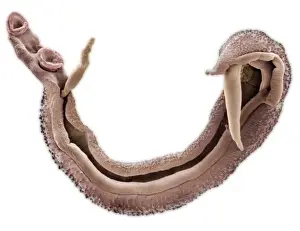
RoundwormScanning electron microscope (SEM) image of a parasitic roundworms head (x 1000 on a standard 9 cm wide print)
Moth eggScanning electron microscope (SEM) image of a moth egg (x 90). The caterpillar emerges by chewing through the shell
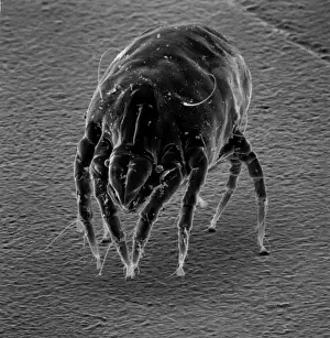
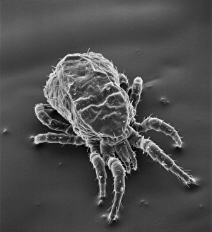
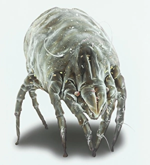
Dermatophagoides sp. dust miteScanning electron microscope image of a dust mite. Dust mites are secondary to pollen as a cause for allergies and they live in bedding, soft furniture and carpets
Sarcoptes scabiei, scabies miteScanning electron microscope image of an itch or scabies mite, a parasite that infests a wide variety of mammalian hosts including man
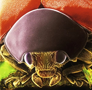
Coccinella sp. black spotted ladybirdScanning electron microscope image showing the head of a black spotted ladybird (x 9 on a standard 9 cm wide print). This image has been coloured artifically by computer
Emiliania huxleyi coccolithophores collected from a bloom in the SW Approaches to the English Channel in June 2004. Date: 2004
Sea saltA scanning electron microscope (SEM) image of sea salt, artificially coloured by computer
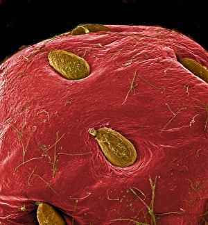
Fragaria sp. strawberryA scanning electron microscope (SEM) image of a strawberry (Fragaria sp.), artificially coloured by computer
Tyrophagus casei, cheese miteScanning electron microscope image of a cheese mite (x 170). These creatures are generally considered to be a pest, however they are added to Altenburger cheese to give it flavour
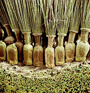
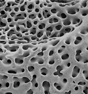
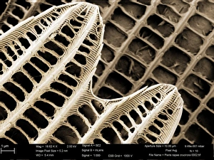
SEM of echinoderm steroemA SEM of an example of stereom of an echinoderm (phylum which consists of 5 classes including starfish). Stereom is the structure formed by the fine networks of calcium carbonate which constitute
Dermanyssus gallinae, red or poultry miteScanning electron microscope image of the red or poutry mite. Adults appear red when engorged with blood, but otherwise are black, grey or white. Females are about 1mm long
Phthiracarus sp. box mite or armadillo miteScanning electron microscope (SEM) image of a box mite, showing how the body has fused into one single segment
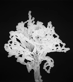
Ceratodon purpureus, ceratodon moss spore capsuleScanning electron microscope (SEM) image of a ceratodon moss spore capsule (x 650 on a standard 9 cm wide print)
ForaminiferScanning electron microscope (SEM) image of a foraminifer - a single celled organism
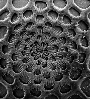
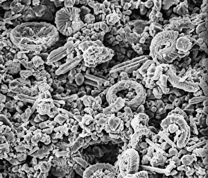
CoccolithScanning electron microscope (SEM) image of a Folkestone chalk surface with Cretaceous coccoliths (x2500 on a standard 9 cm wide print)
Schistosoma nasale, bloodflukeScanning electron microscope image of a parasitic bloodfluke or flatworm. Coloured artifically by computer
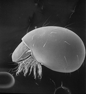
Gyrodactylus, aquatic parasiteScanning electron microscope (SEM) image of a monogenean, Gyrodactylus, a small leech-like parasite on the skin of a salmon (x 600)
Dermatophagoides pteronyssius, dust miteScanning electron microscope image showing a dust mite (x 250 on standard 9cm wide print). This image has been artificially coloured by a computer
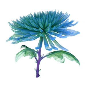
Chrysanthemum, CT scan imageCT Scan image of a Chrysanthemum
Syracosphaera anthosCoccosphere from the Western Mediterranean. False coloured to show the shell is formed of inner and outer layers of coccoliths with very different structure
Papilio palinurus, emerald swallowtailSEM image of an emerald swallowtails wing
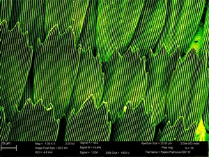
Pieris rapae, small whiteSEM image of the wing of a small white butterfly
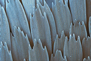
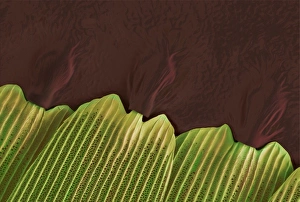
Papilio machaon, old world swallowtailSEM image of Papilio machaon wing
Heliconius doris, doris longwingSEM image of Heliconius doris wing