mail_outline sales@mediastorehouse.com
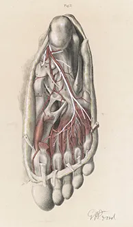
Anatomy / Sole of FootFirst and second stages of the dissection of the sole of the foot
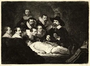
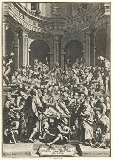
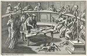
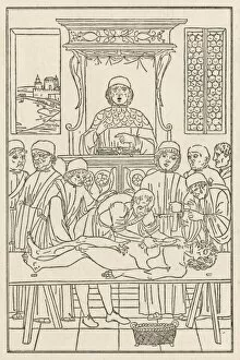
Rembrandts DissectionAn anatomical demonstration
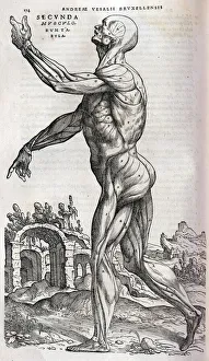
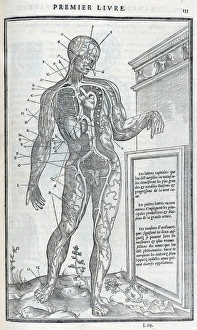
Anatomical drawing of musculature, side viewSecunda Musculorum Tabulae (Anatomical drawing of musculature, side view) Source: Andreae Vesalii Bruxellensis, scholae medicorum Patauinae professoris
Scientist working with a ragworm specimenScientist dissecting a ragworm specimen, held at the Natural History Museum, London
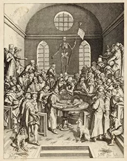
Vesalius DemonstratesVesalius demonstrating
Medical / Anatomy C16Anatomy demonstration. Date: 16th Century
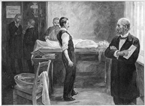
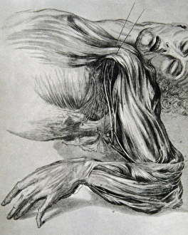
RECOGNITION AT AUTOPSYA surgeon about to perform an autopsy recognises the dead woman lying on the table Date: 1899
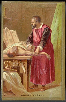
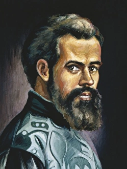
VESALIUS/TRADE CARDANDREAS VESALIUS Belgian anatomist, active in Italy Date: 1514 - 1564
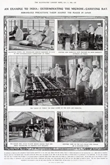
Precautions taken against the plague in Japan, 1908An example to India: exterminating the microbe-carrying rat. Remarkable precautions taken against the plague in Japan. Photographs include sorting
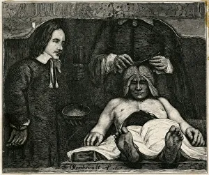
Dr Deijmans Anatomy LessonEngraving after a painting by Rembrandt, showing a physician looking in detail at deceased mans brain
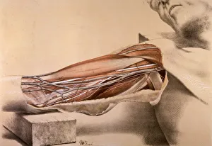
Anatomy, inner arm dissection post-mortemSuperficial view of the arm on the inner side, with the parts undisturbed. Date: 1867
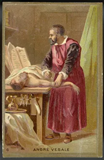
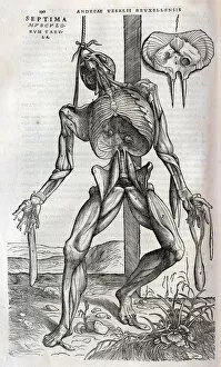
VESALE DISSECTS CORPSEAndre Vesale, an anatomist, born in Brussels, dissects a corpse. Date: circa 1530
Andrea Vesalio. De humani corporis fabricaVesalius, Andreas (1514-1564). Brabantian anatomist, physician, and author of one of the most influential books on human anatomy, De humani corporis fabrica. Oil
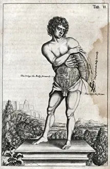
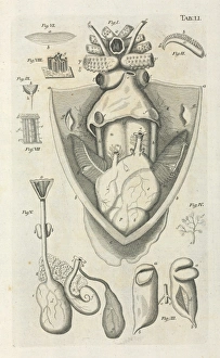
Naked male on a plinth with cross section of stomachTab II. Naked male on a plinth with cross section of stomach Source: Myographia nova, or, A graphical description of all the muscles in the humane body, as they arise in dissection by Browne, John
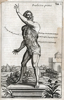
Naked man on plinth with cross section of musclesPraelectio secunda - Tab I. Naked man on plinth with cross section of side and shoulder muscles Source: Myographia nova, or, A graphical description of all the muscles in the humane body
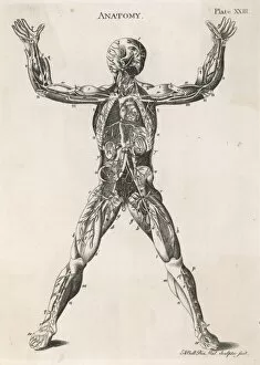
Anatomical drawing of the human bodyAn anatomical drawing of the human body, showing muscles, nerves and internal organs
Drawing from a study of anatomy. Human muscles of the armsHistory of medicine. Drawing from a study of anatomy. Human muscles of the arms. 18th century
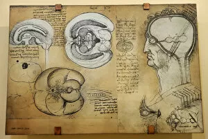
Nervous system Leonardo da Vincis drawing. 15 th century. Leonardo studies the central nervous system in the ox brain an ingenious technique allows him to reproduce the shape of the brain ventricles
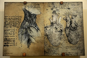
Cardiovascular system Leonardo da vincis drawingCardiovascular systems drawing. In 1513 Leonardo da Vinci to study the heart and the circulatory system through animal dissections. Windsor, Royal Library 19073, 1510-1513
Anatomical drawing of musculatureSeptima Musculorum Tabula (Anatomical drawing of musculature) Source: Andreae Vesalii Bruxellensis, scholae medicorum Patauinae professoris
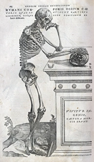
Anatomical drawing of a human skeletonHumani Corporis ossium caeteris quae sustinent partibus liberorum suaque sede positorum (Anatomical drawing of a human skeleton side view) Source: Andreae Vesalii Bruxellensis
Macropoma lewesiensis, an extinct coelacanth fishRestoration of one of the last coelacanth fishes (Macropoma lewesiensis). It lived in the clear water chalk seas of sourthern England 85 mya, and grew to about 60 cm in length
Anatomical Theatre C16Anatomy in the 16th century as depicted in Bartholinus Eustachius works. It looks as though dogs are enjoying the unwanted organs
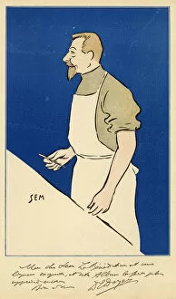
Eugene-Louis Doyen / SemEUGENE-LOUIS DOYEN French medical Date: 1859 - 1916
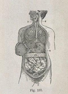
VALUEHuman Anatomy. Digestive system. Engraving
Anatomical drawings; dissection Source: La dissection des parties du corps humain divisee en trois livres / faictz par Charles Estienne
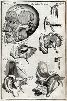
Parts of the headTab VII - Praelectio secunda. A head and other diagrams showing parts of the head Source: Myographia nova, or, A graphical description of all the muscles in the humane body
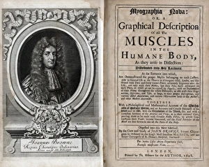
Title page - frontispiece - Portrait of John BrowneTitle page and frontispiece of work - Portrait of John Browne Source: Myographia nova, or, A graphical description of all the muscles in the humane body, as they arise in dissection by Browne, John
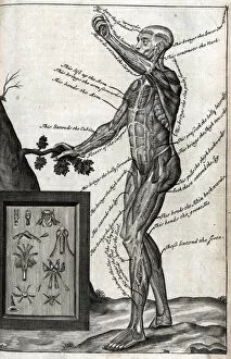
View of male from side Source: Myographia nova, or, A graphical description of all the muscles in the humane body, as they arise in dissection by Browne, John, 1642-ca. 1700
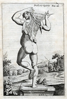
Male on plinth viewed from rearPraelectio Quarta - Tab XV. Male on plinth viewed from rear, with cross section of back muscles Source: Myographia nova, or, A graphical description of all the muscles in the humane body
Cuttlefish dissection drawingTable LI, taken from Bibel der Natur byJan Swammerdamm
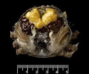
Dissected male Eriocheir sinensis, Chinese mitten crabA dissected male mitten crab (eriocheir sinensis), showing the ripening testes. Specimen was collected from the River Thames
Dissected female Eriocheir sinensis, Chinese mitten crabA dissected female mitten crab (eriocheir sinensis), showing the ripening ovaries. Specimen was collected from the River Thames
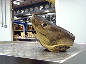
Lamna nasus, porbeagle sharkSpecimen of a porbeagle sharks head on the dissection table outside the Tank Room of the Darwin Centre, at the Natural History Museum, London
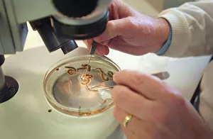
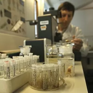
Scientist at work at The Natural History Museum, LondonAccurate identification of copepod crustaceans is a time consuming task for specialists, requiring meticulous sorting, dissection, and the use of high resolution microscopy
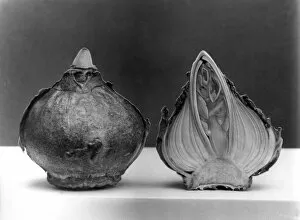
Hyacinth BulbA hyacinth bulb cut in two, with a section clearly showing the leaves and flower inside the bulb. Date: 1960s
First Dissection PicThis is said to be the earliest depiction of an anatomical dissection
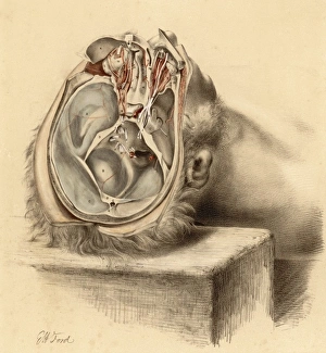
Anatomy / Base of SkullBase of the skull and second views of the orbit
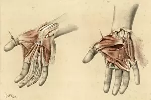
Anatomy / Hands / 1867Superficial and deep views of the palm of the hand
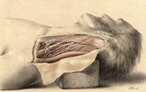
Anatomy / Side of NeckThe anatomy of the side of the neck behind the sternomastoid muscle
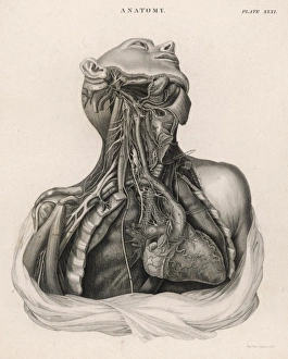
Dissection of upper torso, showing the heartA dissection of the upper torso of a human body, showing the heart, lungs, veins and arteries
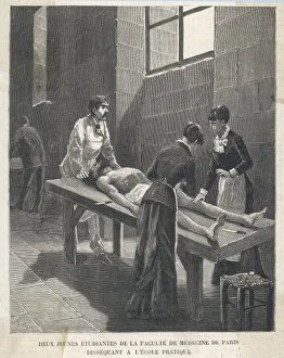
Students Dissecting 1880Young female students at the Faculty of Medicine in Paris dissect a body as part of their practical work