mail_outline sales@mediastorehouse.com
Choose a picture from our Micro Photography Collection for your Wall Art and Photo Gifts
47 items
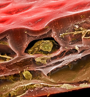
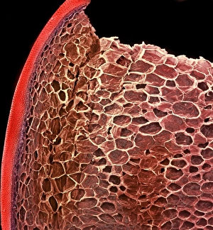
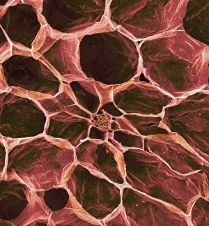
Human cellIllustration of a highly magnified section through a human cell. Page 8 from Human Biology, 1977
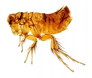
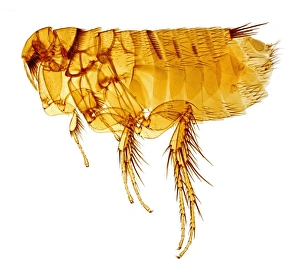
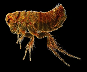
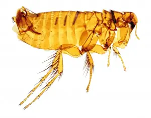
Tunga penetrans, chigoe fleaThis species of flea is commonly known as a jigger, chigoe or sand-flea
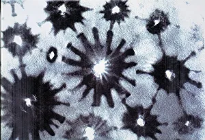
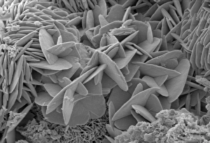
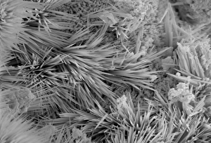
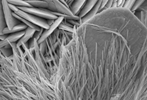
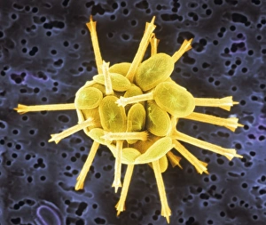
Crysotile asbestosScanning electron micrograph of 5-Fold symmetry in crysotile asbestos. Magnification on the 5 x4 transparency = X 600, 000
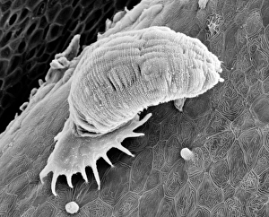
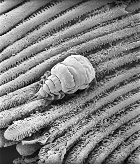
Gyrodactylus, aquatic parasiteScanning electron microscope (SEM) image of a monogenean, Gyrodactylus, a small leech-like parasite on the skin of a salmon (x 600)
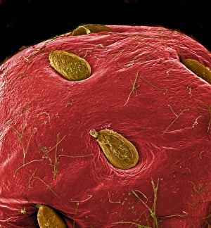
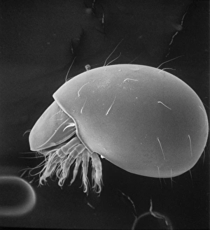
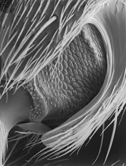
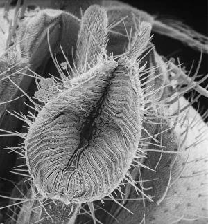
Moth eggScanning electron microscope (SEM) image of a moth egg (x 90). The caterpillar emerges by chewing through the shell
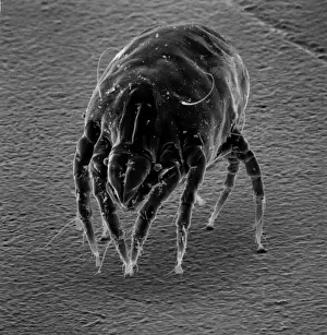
Dermatophagoides sp. dust miteScanning electron microscope image of a dust mite. Dust mites are secondary to pollen as a cause for allergies and they live in bedding, soft furniture and carpets
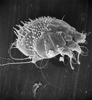
Sarcoptes scabiei, scabies miteScanning electron microscope image of an itch or scabies mite, a parasite that infests a wide variety of mammalian hosts including man
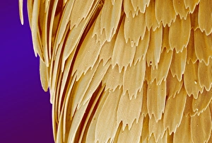
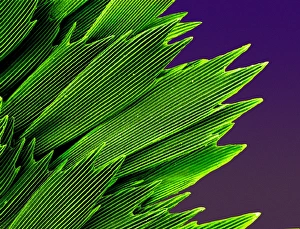
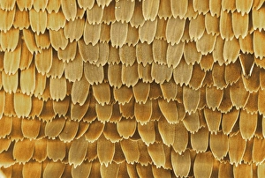
Papilio machaon, old world swallowtailSEM image of a Papilio machaon wing
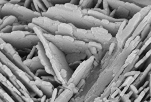
Malachite
Malachite comprises of (copper carbonate hydroxide). Malachite has distinctive green banding and belongs to the carbonate class
Papilio machaon, old world swallowtailSEM image of Papilio machaon wing
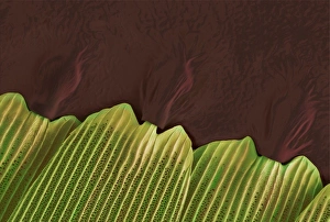
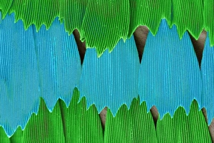
Papilio palinurus, emerald swallowtailSEM image of an emerald swallowtails wing
Papilio palinurus, emerald swallowtailSEM image of an emerald swallowtail wing
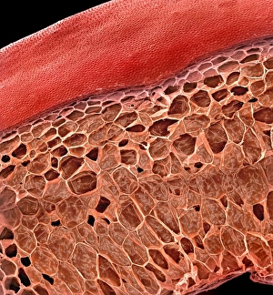
Solanum sp. tomatoA scanning electron microscope (SEM) image of a tomato (Solanum sp.), artificially coloured by computer
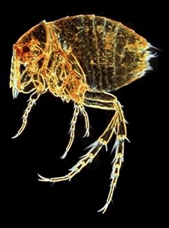
Hystrichopsylla talpae talpae, mole fleaA macro photograph of the largest flea in the UK, the mole flea (Hystrichopsylla talpae talpae), which is common on small mammals throughout the UK
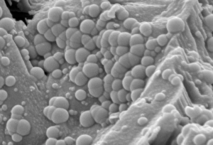
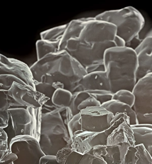
Table saltA scanning electron microscope (SEM) image of table salt, artificially coloured by computer
OatsA scanning electron microscope (SEM) image of oats, artificially coloured by computer
Fragaria sp. strawberryA scanning electron microscope (SEM) image of a strawberry (Fragaria sp.), artificially coloured by computer
Solanum sp. tomato
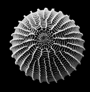
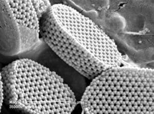
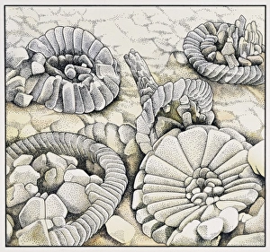
CoccolithsScanning electron microscope (SEM) image of coccoliths, these are the limestone scales surrounding the marine phytoplankton coccolithophores
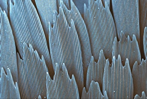
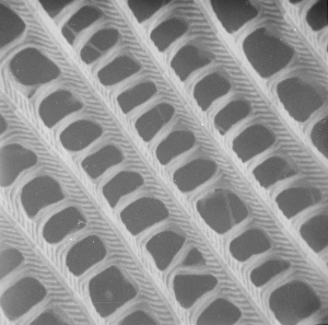
Butterfly wing scale (part)
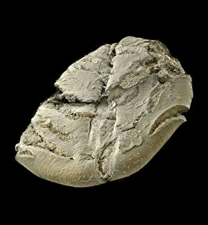
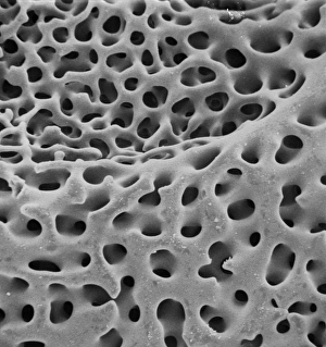
SEM of echinoderm steroemA SEM of an example of stereom of an echinoderm (phylum which consists of 5 classes including starfish). Stereom is the structure formed by the fine networks of calcium carbonate which constitute
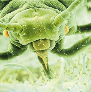
Aphis fabae, black bean aphidScanning electron microscope image showing a frontal view of a black bean aphid on leaf (x100). Aphids or plant lice are small, plant-sucking insects
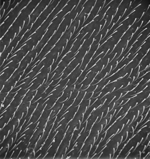
Calliphora vicina, blowfly or blue bottleScanning electron microscope (SEM) image of a blowflys wing
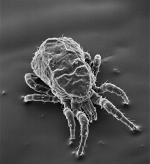
Dermanyssus gallinae, red or poultry miteScanning electron microscope image of the red or poutry mite. Adults appear red when engorged with blood, but otherwise are black, grey or white. Females are about 1mm long
Phthiracarus sp. box mite or armadillo miteScanning electron microscope (SEM) image of a box mite, showing how the body has fused into one single segment
Fagus sylvatica, European beech pollenScanning electron microscope picture (X1500) showing a pollen grain as seen from the side. The image shows one of the three laterally-placed aperture furrows with a small pore in the centre
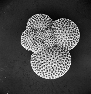
ForaminiferScanning electron microscope (SEM) image of a foraminifer - a single celled organism
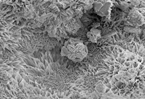
Calyptrolithophora papillifera, holococcolithAn SEM of a holococcolith, a nano-fossil, with flat top
Lasius niger, black garden antScanning electron microscope (SEM) of a black ant leg. Widespread and common in a range of habitats but perhaps most familiar in gardens where nests are formed under paving stones and brickwork
Calliphora vicina, blowfly or bluebottleScanning electron microscope (SEM) image of a blowfly proboscis (x 85). This specialised mouth-part is used to squirt digestive enzymes onto the food
Papilio machaon, old world swallowtailSEM image of the wing of Papilio machaon
Coccoliths magnified a thousand timesAn illustration of Coccoliths magnified a thousand times. Coccoliths are micro-fossils and feature heavily in the composition of chalk
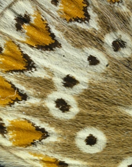
Polyommatus icarus, common bluePhotograph of a mounted specimen of the common blue, highly magnified scales from the hindwing underside of the female butterfly
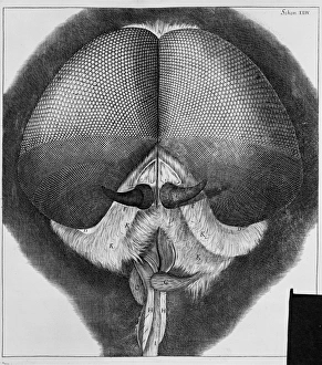
Eye of a flyPage 201. From Micrographia or some Physiological description of minute bodies made by magnifying glasses, 1665 by Robert Hooke