mail_outline sales@mediastorehouse.com
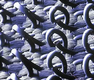
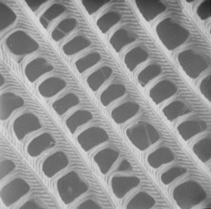
VelcroA trademarked name for a fastening tape made up of a strip of nylon with a surface of minute hooks, that fasten to another strip with a surface of uncut pile. A SEM image
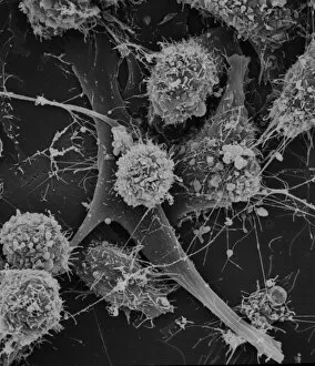
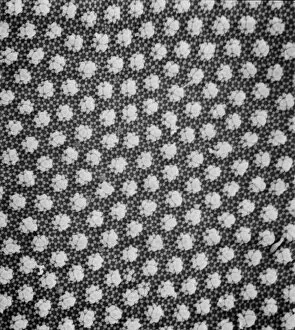
Cells on glassScanning electron microscope (SEM) image of cells on glass (x 2K)
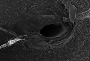
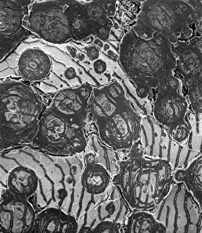
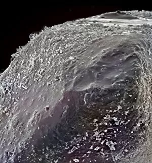
Volcanic glass, Peles hairScanning electron microscope image of a sample of volcanic glass from Mt. Pele, produced to evaluate different types of laser in Laser Ablation Inductively Coupled Plasma Mass Spectrometry
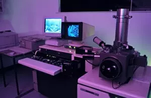
Variable pressure scanning electron microscopeThis electron microscope allows the imaging of samples without any preparation
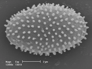
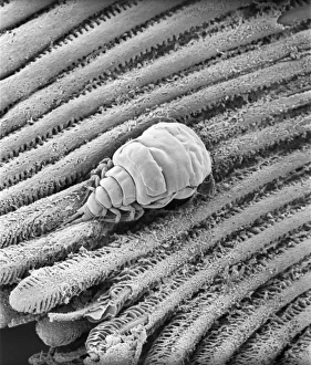
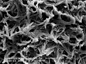
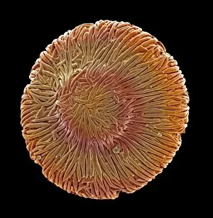
Myxomycetes, plasmodial slime mouldScanning electron microscope image of a plasmodial slime mould spore (x12000). This mould spends most of its life as a single cell; when they reproduce they form a slug-like blob that can travel
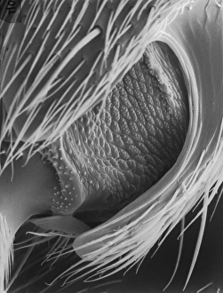
Collembola ocelli, springtailScanning electron microscope image of the springtail with simple eyes (x 1.2K)
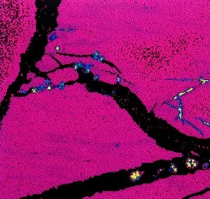
Copper in unspecified mineralScanning electron microscope image of an elemental map showing the distribution of copper (Cu) in mineral samples
OstracodScanning electron microscope image of an ostracod, an arthropod where the body is enclosed in a carapace (x 220)
Collembola sp. springtailScanning electron microscope image of a springtail showing the characteristic pattern on the cuticle surface (x 3.5K)
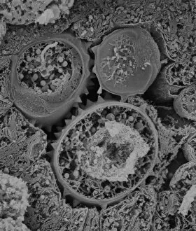
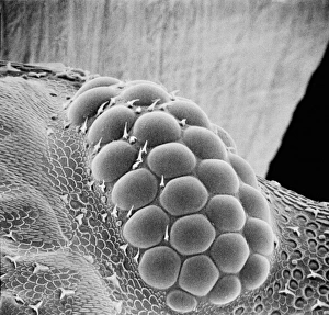
Fractured pollen grainScanning electron microscope (SEM) image showing a fractured pollen grain
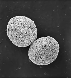
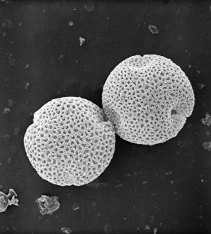
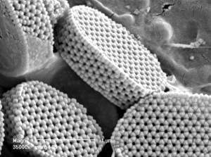
Populus nigra, lombardy or black poplar pollenScanning electron microscope image (x 1500) of black poplar pollen grains showing a characteristic granular surface ornamentation and no apertures (inaperturate)
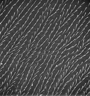
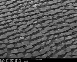
Butterfly wing scale (part)
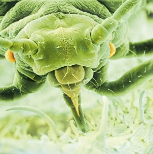
Aphis fabae, black bean aphidScanning electron microscope image showing a frontal view of a black bean aphid on leaf (x100). Aphids or plant lice are small, plant-sucking insects
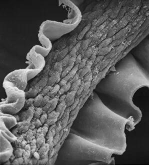
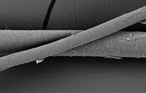
Human hairScanning electron microscope (SEM) image showing a human hair with the cuticle reflexed
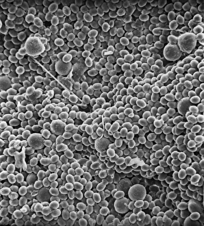
Coccolithus pelagicusCoccosphere of Coccolithus pelagicus, a common cold water coccolithophore. Collected from the British Continental shelf, North West of Scotland. Specimen diameter 15m. False-coloured SEM image
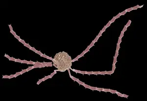
Ophiaster formosusA coccolithophore with long appendages formed of strings of highly modified coccoliths. Collected from the West Pacific. Specimen diameter 50m. False-coloured SEM image
Lasius niger, black garden ant
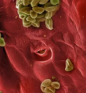
Calliphora vicina, blowfly or blue bottleScanning electron microscope (SEM) image of a blowflys wing
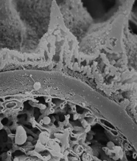
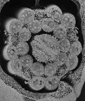
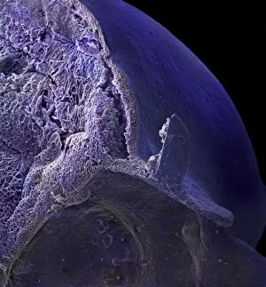
Fractured antherScanning electron microscope (SEM) image showing a fractured anther, otherwise known as the sac, which contains the pollen in the male sex organs (stamens)
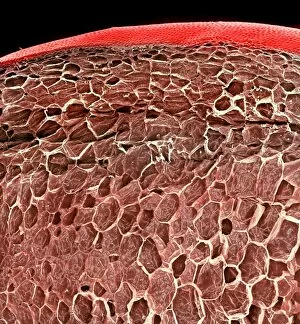
Asteraceae, daisyScanning electron microscope image of the fractured surface of an anther showing a developing pollen grain from a member of the daisy or Asteraceae family ( X 3000)
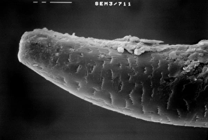
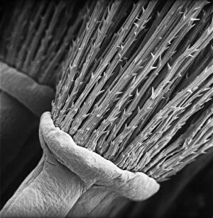
Lumbricus terrestris, earthwormScanning electron microscope (SEM) image showing the chaeta/setae - involved in the locomotion on an earthworm
Taraxacum officinale, dandelionScanning electron microscope (SEM) image of a dandelion (x 80)
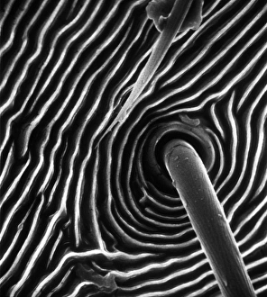
Spider trichobothrium hairScanning electron microscope (SEM) image of the base of a trichobothrium hair (x 1, 000). The hair is an air-movement sensor extending from the pit in the cuticle of a spiders leg
Fagus sylvatica, European beech pollenScanning electron microscope picture (X1500) showing a pollen grain as seen from the side. The image shows one of the three laterally-placed aperture furrows with a small pore in the centre
Fraxinus excelsior, weeping ash pollenScanning electron microscope picture (x 1500) of ash pollen grains from above, with three furrowed apertures (trizonocolporate)
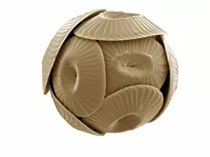
Calyptrolithophora papillifera, holococcolithAn SEM of a holococcolith, a nano-fossil, with flat top
A bryozoan colonyScanning electron microscope image displayed on the glass screens in the Darwin Centre, at the Natural History Museum, London
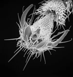
Cystopteris diaphana, diaphanous bladder fernAn SEM showing a close-up of the spiny-lacunar surface of the diaphanous bladder fern (Cystopteris diaphana) spore. Photographed using Philips XL30 SEM
Hair of the DogA scanning electron micrograph (SEM) of a dog hair
Mustelus mustelus, smoothhound sharkA Scanning Electron Microscope image of smoothhound shark skin. The skin is covered with tiny teeth called dermal denticles
Fragaria sp. strawberryA scanning electron microscope (SEM) image of a strawberry (Fragaria sp.), artificially coloured by computer
Sugar grainsA scanning electron microscope (SEM) image of sugar grains, artificially coloured by computer
Vitis sp. white grapeA scanning electron microscope (SEM) image of a white grape (Vitis sp.), artificially coloured by computer
Browallia speciosa, amethystA pollen grain of the Browallia speciosa (polar view) from the family Solanaceae, the tomato family
Vaccinium sp. blueberryA scanning electron microscope (SEM) image of a blueberry (Vaccinium sp.), artificially coloured by computer
Solanum sp. tomatoA scanning electron microscope (SEM) image of a tomato (Solanum sp.), artificially coloured by computer
Isurus oxyrinchus, mako sharkScanning Electron Microscope image of mako shark skin
Lasius niger, black garden antScanning electron microscope (SEM) of a black ant leg. Widespread and common in a range of habitats but perhaps most familiar in gardens where nests are formed under paving stones and brickwork
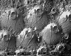
Porcellio sccaber, woodlouseScanning electron microscope (SEM) image showing all the units that make up the compound eye of a woodlouse
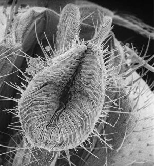
Calliphora vicina, blowfly or bluebottleScanning electron microscope (SEM) image of a blowfly proboscis (x 85). This specialised mouth-part is used to squirt digestive enzymes onto the food
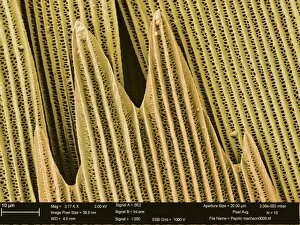
Papilio machaon, old world swallowtailSEM image of a Papilio machaon wing
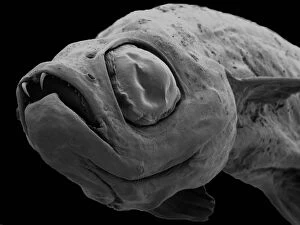
Danionella dracula, minnowSEM image of the Danionella dracula. This tiny 17mm fish has evolved many unique and unusual characteristics, the most spectacular of which are jaw modifications that resemble true teeth
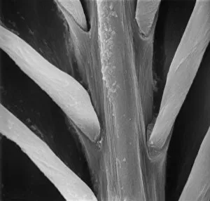
Feather detail
Pollen on beeScanning electron microscope (SEM) image of pollen on a bee. If the plant depends on animals for pollination, the pollen will be relatively large and sticky
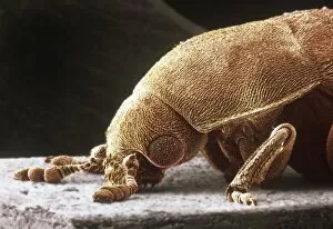
Dermestes lardarius, larder beetleScanning electron microscope image of a larder beetle (x22). These beetles are important for the damage they do, mainly through feeding on animal matter. Coloured artificially by computer