mail_outline sales@mediastorehouse.com
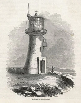
Old Hartlepool Lighthouse, north east EnglandView of the old Hartlepool Lighthouse, north east England (County Durham), completed in 1847. A man with a telescope scans the horizon
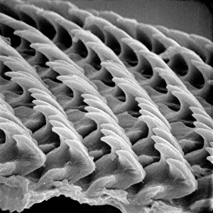
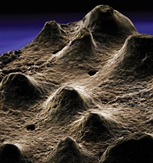
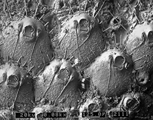
Snail teeth
Plasmodium sp. malarial parasiteScanning electron microscope image of a malarial protozoal parasite. The parasite requires the anopheles mosquito to complete its life cycle
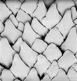
Scyliorhinus canicula, dogfishScanning electron microscope (SEM) image of the scales of a dogfish (x 40)
AspergillusAn SEM image of aspergillus in spore production (x 815 on a standard 9 cm wide print). The moulds are common in the northern hemisphere and some cause disease in humans and animals
The anus of a bot flyScanning electron microscope image of the anus of a bot fly. Image on display in the Darwin Centre at the Natural History Museum, London
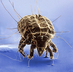
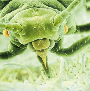
Tyrophagus casei, cheese miteScanning electron microscope image of a cheese mite (x 170). These creatures are generally considered to be a pest, however they are added to Altenburger cheese to give it flavour
Ceratodon purpureus, ceratodon moss spore capsuleScanning electron microscope (SEM) image of a ceratodon moss spore capsule (x 650 on a standard 9 cm wide print)
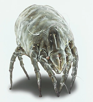
Dermatophagoides pteronyssius, dust miteScanning electron microscope image showing a dust mite (x 250 on standard 9cm wide print). This image has been artificially coloured by a computer
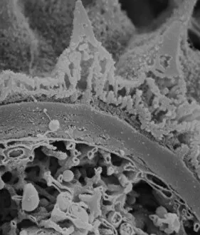
Dinosaur eggshellScanning electron microscope image on display in the Darwin Centre
Ventral surface of a mite from the prostigmatic speciesScanning electron microscope image displayed on the glass screens in the Darwin Centre, at the Natural History Museum, London
Diplopoda sp. plate millipedeScanning electron microscope image of a lateral view of the head of a plate millipede. Image displayed on the glass screens in the Darwin Centre, at the Natural History Museum, London
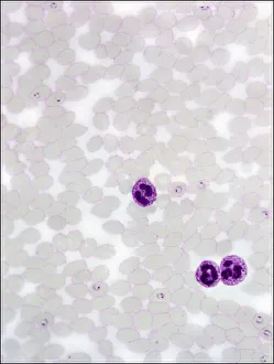
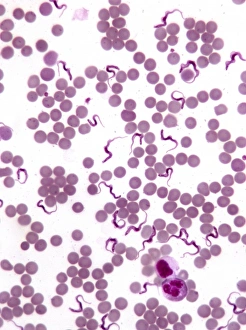
TrypanosomesScanning electron microscope image showing a trypanosoma blood smear. They have proved to be of great interest as they have evolved very differently to other better studied organisms
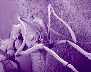
Atta cethalotes, leaf-cutter antScanning electron microscope image of a leaf-cutter ant displayed in the Darwin Centre, at the Natural History Museum, London
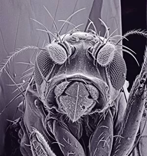
Small fly, species unknownScanning electron microscope (SEM) of a fly head. Image displayed on the glass screens in the Darwin Centre, at the Natural History Museum, London
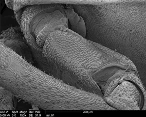
Woodlouse antennaScanning Electron Microscope (SEM) image of woodlouse antenna
Amphitetras, diatomScanning electron microscope (SEM) image showing the diatom Amphitetras with its ornate silica shell (x5000 on a standard 9 cm wide print). Coloured artificially by computer
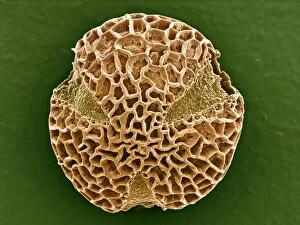
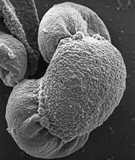
Leptoglossis ferreyraeiA pollen grain of Leptoglossis ferreyraei (polar view) from the family Solanaceae, the tomato family
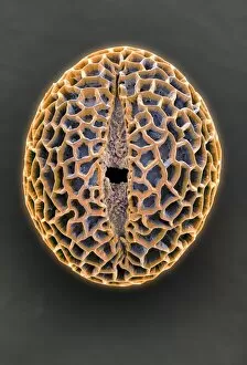
Leptoglossis lomanaA pollen grain of the Leptoglossis lomana (polar view) from the family Solanacea, the tomato family
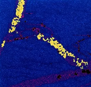
Gold in unspecified mineralScanning electron microscope image of an elemental map showing the distribution of gold (Au) in mineral samples
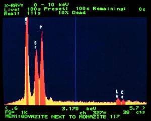
GoyaziteScanning electron microscope image of the energy-dispersive X-ray spectrum of the mineral goyazite, obtained using Link AN10000 analysis system
VelcroA trademarked name for a fastening tape made up of a strip of nylon with a surface of minute hooks, that fasten to another strip with a surface of uncut pile. A SEM image
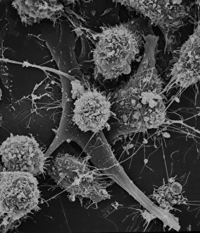
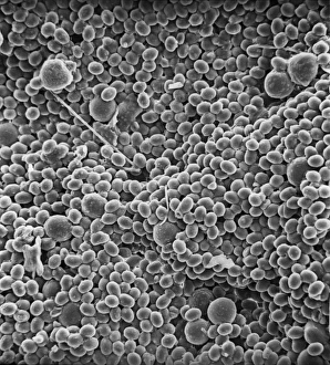
Cells on glassScanning electron microscope (SEM) image of cells on glass (x 2K)
Copper in unspecified mineralScanning electron microscope image of an elemental map showing the distribution of copper (Cu) in mineral samples
Fractured pollen grainScanning electron microscope (SEM) image showing a fractured pollen grain
Aphis fabae, black bean aphidScanning electron microscope image showing a frontal view of a black bean aphid on leaf (x100). Aphids or plant lice are small, plant-sucking insects
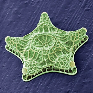
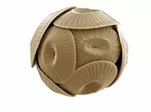
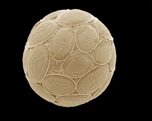
Coccolithus pelagicusCoccosphere of Coccolithus pelagicus, a common cold water coccolithophore. Collected from the British Continental shelf, North West of Scotland. Specimen diameter 15m. False-coloured SEM image
Florisphaera profundaA coccolithophore with highly modified, plate-like coccoliths. This is a very common deep dwelleing species, typically living at about 100-150m depth in the water column
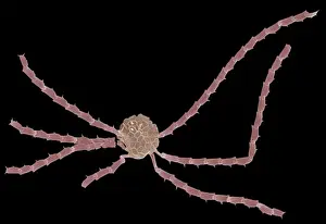
Ophiaster formosusA coccolithophore with long appendages formed of strings of highly modified coccoliths. Collected from the West Pacific. Specimen diameter 50m. False-coloured SEM image
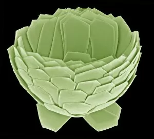
Pontosphaera japonica. A coccolithophore with relatively large, flat, coccoliths. Collected from off Hawaii. Specimen diameter 22m. False-coloured SEM image
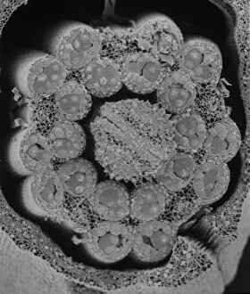
Fractured antherScanning electron microscope (SEM) image showing a fractured anther, otherwise known as the sac, which contains the pollen in the male sex organs (stamens)
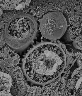
Asteraceae, daisyScanning electron microscope image of the fractured surface of an anther showing a developing pollen grain from a member of the daisy or Asteraceae family ( X 3000)
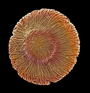
Taraxacum officinale, dandelionScanning electron microscope (SEM) image of a dandelion (x 80)
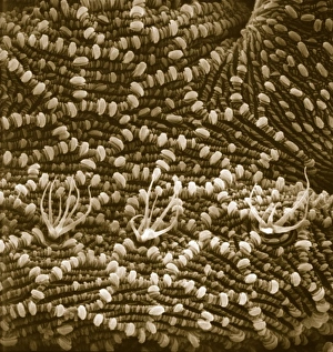
A bryozoan colonyScanning electron microscope image displayed on the glass screens in the Darwin Centre, at the Natural History Museum, London
Browallia speciosa, amethystA pollen grain of the Browallia speciosa (polar view) from the family Solanaceae, the tomato family
Pollen on beeScanning electron microscope (SEM) image of pollen on a bee. If the plant depends on animals for pollination, the pollen will be relatively large and sticky
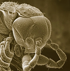
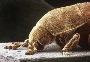
Dermestes lardarius, larder beetleScanning electron microscope image of a larder beetle (x22). These beetles are important for the damage they do, mainly through feeding on animal matter. Coloured artificially by computer
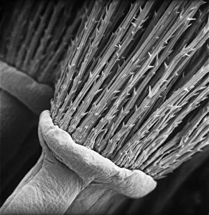
Pinus sylvestris, scots pineScanning electron microscope (SEM) image showing a pollen grain from a scots pine. Note the air bladders that help it to float through the air (x 1500 on a standard 9 cm wide print)
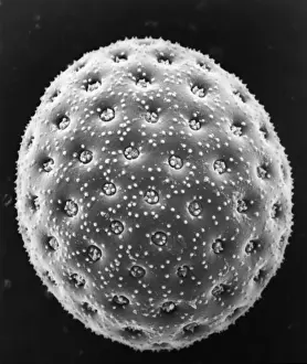
Chenopodium album, goosefootScanning electron microscope image of a pollen grain from a member of the goosefoot family (x 3000 on a standard 9 cm wide print)
Relatives and friends scanning the fateful lists at the CanaScanning the fateful lists for news. Relatives and friends of passengers on the Empress of Ireland making inquiries at the Canadian Pacific Railway offices in Cockspur Steet