mail_outline sales@mediastorehouse.com
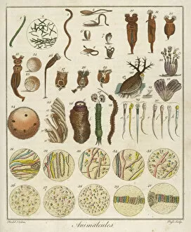
Under the Microscope / C18Animacules : microscopic creatures as seen under a microscope; the last two rows are human sperm
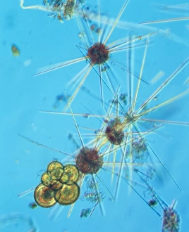
Microscopic Marine LifeVarious forms of microscopic marine life, described by Sibly as Animalcules Date: 1794
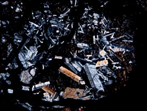
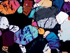
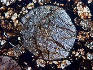
Microscope image of the Pasamonte eucriteMicroscopic image of the Pasamonte eucrite showing a basaltic texture. Field of view is 2.5mm across
Microscopic views of human spermatozoa in semenMicroscopic views of human spermatozoa. View of the animalculae or organic particles in the semen. Handcoloured copperplate engraving by J
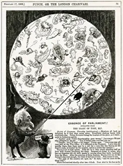
Cartoon, Essence of Parliament (MPs)Cartoon, Essence of Parliament -- Mr Punch shows members of the House of Commons as microscopic bugs in a petri dish. 1883
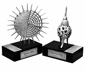
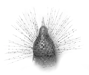
Radiolaria modelsModels of two radiolaria made in papier mache by Vaclav Fric
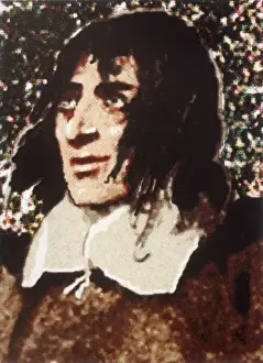
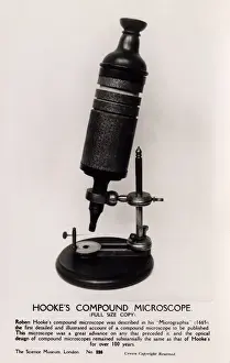
ROBERT HOOKE 1635 - 1703ROBERT HOOKE English scientist. Author of Micrographia (1665), in which he published results of his microscopic investigations
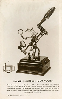
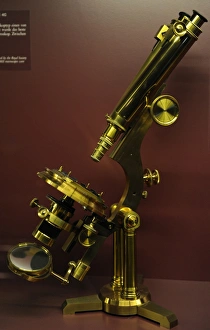
Adams Universal Microscope - made by George Adams Snr. about 1755 for King George III, while he was still Prince of Wales
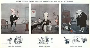
More Types From Harley Street by H M Bateman. Skin - The Microscopic. The one with the magnifying glass and the test tubes. Brain - The Genius. He knows all about the mind. Nerve - The Intense
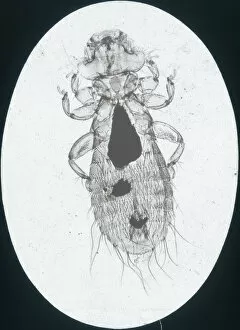
X-Ray - Microscopic x-ray view of a small head louse. Part of Box 165 Boswell Collection - X-Rays. Date: circa 1900
Binocular microscope large Best. London, around 1890Binocular microscope large Best. Signed: R. & J. Beck Ltd London 19901. London, around 1890. The Large Best microscope was the top product of R. and J
Changeable cercaria, Cercaria mutabilis.. Handcolored copperplate zoological engraving from George Shaw and Frederick Nodders The Naturalists Miscellany, 1792
Common moss, Phascum cuspidatum, and pond algae.. Magnified image of common moss, Phascum cuspidatum 1, and microscopic enlargment of pond algae, Hydrodictyon utriculatum 2
Robert Hookes MicroscopeFull-size copy of Robert Hookes Compound Microscope - held at the Science Museum, London. Hooke, an English natural philosopher, architect and polymath (1635-1703) - author of Micrographia (1665)
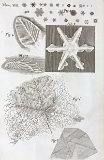
Schem VIII from Robert Hookes MicrographiaSchem VIII located between pages 88 & 89 in Micrographia: or Some physical descriptions of minute bodies made by magnifying glasses, with observations and enquiries thereupon
Radiolarian modelGalls model of radiolarian by Blaschka, held at the Natural History Museum, London
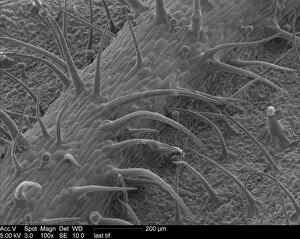
Pelargonium sp. geraniumScanning Electron Microscope image of a pelaronium leaf
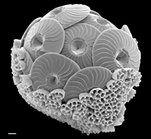
CoccolithsScanning electron microscope (SEM) image of coccoliths, these are the limestone scales surrounding the marine phytoplankton coccolithophores
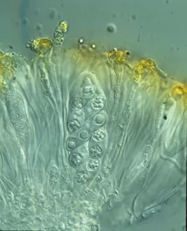
Xanthoria parietina, lichenShown here is a maritime sunburst lichen. A photograph of the ascus containing eight ascopores
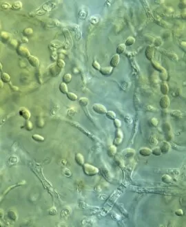
Nostoc spA cyanobacterium. Members of this genus occur frequently in lichens, especially those in wet habitats
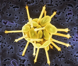
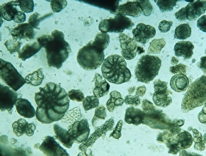
AcanthowetraA photograph of a foraminifera found in the Indian Ocean
Foraminiferan remains from the White Cliffs of Dover, U.K. The cliffs are made up of unimaginable numbers of chalky shells of long dead marine animals
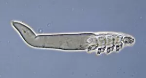
Demodex uncii, follicle miteA follicle mite magnified X600. The follicle mite is a worm-like microscopic mite which lives in the follicles and sebaceous gland of most humans
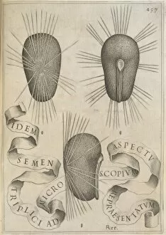
Hibiscus seedsAn illustration of three hibiscus seeds as seen through a microscope, from page 497 of Flora, overo Cultura di Fiori (1638) by Battista Giovanni Ferrari (1582-1655)
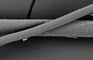
Hair of the DogA scanning electron micrograph (SEM) of a dog hair
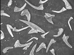
Conodonts, tooth like fossilsThese microscopic tooth like fossils are from the Ordovician period of the Ludlow area, Shropshire, UK about 420m yrs old (Magnification x 3.8)
Microscope image of the Johnstown diogenite. Diogenites are coarse grained and composed primarily of one mineral, pyroxene. Field of view is 2.5mm across
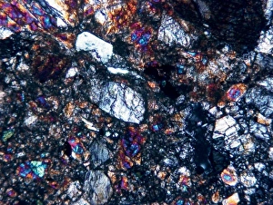
Microscope image of the Zagami shergottite. The fractures in the pyroxene mineral grains and the paler patches of glass show that the rock has been shocked. Field of view is 5mm
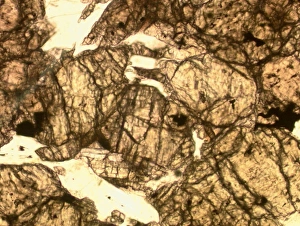
Microscope image of the Lodran meteorite. This meteorite is the type specimen of the Lodranite meteorites. The lodranites are related to the acaplucoites but are more course-grained
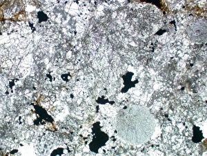
Optical microscope image of the Barwell (Type 6) chondrite. This meteorite has experienced a significant amount of heating
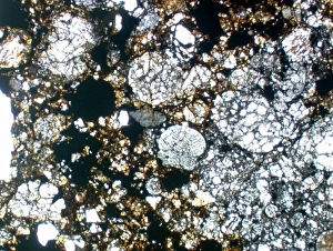
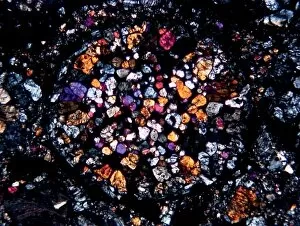
Optical microscope image of the Parnallee (Type 3) chondriteAn optical microscope image of the Parnallee (Type 3) chondrite that has experienced little heating. The chondrules are clear and well-defined. The field of view is 5mm
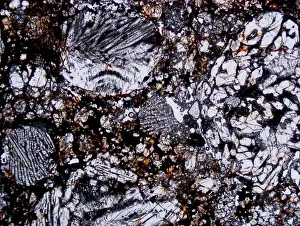
Textures of different chondrule types in the Etihudna (L4) ordinary chondrite (field of view 4mm)
Porphyritic olivine and pyroxene chondruleMicroscopic image of a porphyritic olivine and pyroxene chondrule from the Palmyra (L3) ordinary chondrite (the chondrule is about 1.8mm across)
Radial pyroxene chondruleMicroscope image of a radial pyroxene chondrule from the ALH 88036 (H3.4) ordinary chondrite. The chondrule is about 2mm across
Calcidiscus leptoporus and Syracolithus quadriperforatus, coIn this scanning electron micrograph, the transition of a life-cycle stage in Calcidiscus is shown from the outer cover to the inner layer. Specimen taken from W. Mediterranean
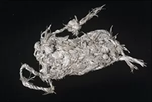
Asbestos purseBenjamin Franklins Asbestos purse. A crudely plaited purse made from tremolite asbestos. From the Hans Sloane collection. Asbestos is formed of microscopically fibrous crystals
German Medical StudentsA group of seated (mostly female) medical students, watching a microscopic projector of what appears to be an insect. Date: 1930s
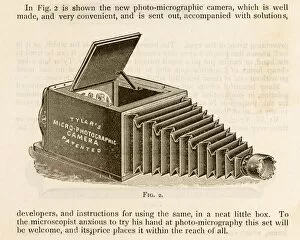
Photo-MicrographiccameraTylers photo-micrographic camera, for photographing microscopic subjects
A Drop of London WaterTHE WONDERS OF A LONDON WATER DROP A water drop as it would be seen under the Molecular Magnifier. It is home to many hideous microscopic creatures
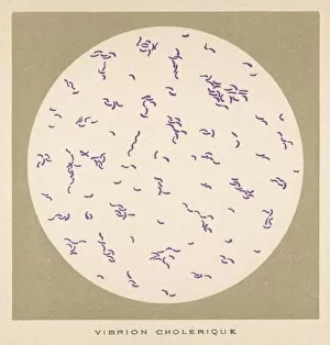
Cholera VibrioVibrio (or vibrion) of cholera, discovered by Koch, 1883
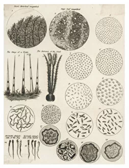
Microscopic ObjectsA variety of living and non- living objects magnified through a microscope