mail_outline sales@mediastorehouse.com
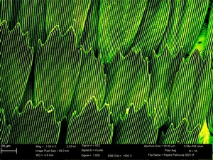
Papilio palinurus, emerald swallowtailSEM image of an emerald swallowtails wing
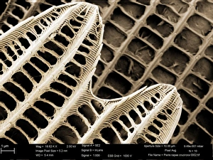
Pieris rapae, small whiteSEM image of the wing of a small white butterfly
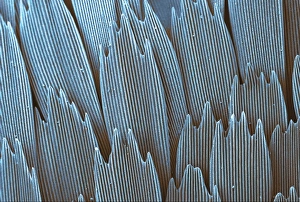
Papilio machaon, old world swallowtailSEM image of Papilio machaon wing
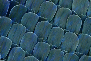
Heliconius doris, doris longwingSEM image of Heliconius doris wing
Malachite comprises of (copper carbonate hydroxide). Malachite has distinctive green banding and belongs to the carbonate class
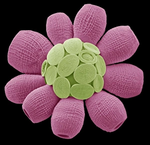
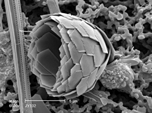
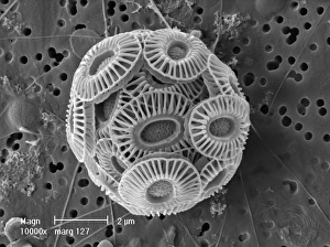
Scyphosphaera apsteinii. SEM image of an equatorial coccolith
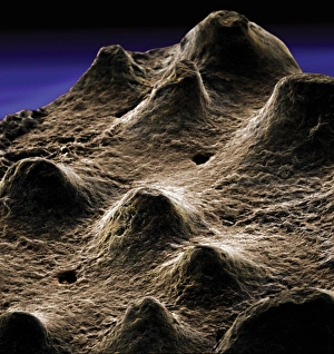
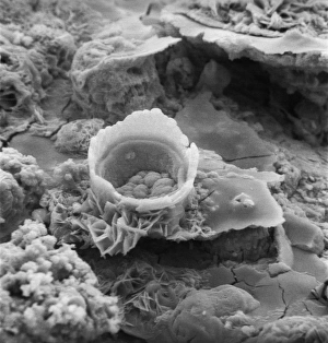
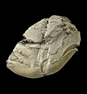
Dinosaur eggshellScanning electron microscope image on display in the Darwin Centre
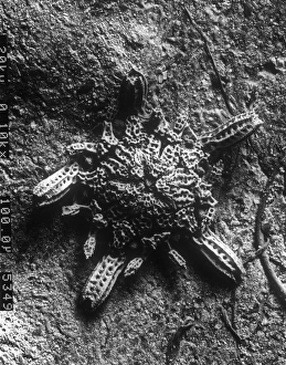
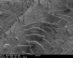
Ventral surface of a mite from the prostigmatic speciesScanning electron microscope image displayed on the glass screens in the Darwin Centre, at the Natural History Museum, London
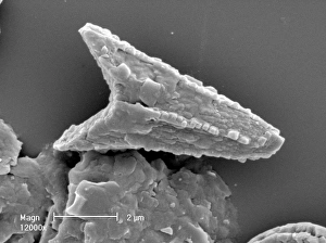
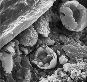
Visbyshaera oligofurcata, acritarchScanning electron microscope image of a microfossil belonging to a group of marine phytoplanktonic organisms known as acritarchs that teemed in Silurian seas about 415 Ma ago
Oestridae, botfly larvaScanning electron microscope image of a botfly larva. They are parasites feeding on skin in the case of warble flies, nostrils in the flies that affect sheep and deer
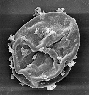
Difflugia CoronaFreshwater Testate Amoebae. Magnification x 450
Lycopod
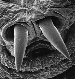
Diplopoda sp. plate millipedeScanning electron microscope image of a lateral view of the head of a plate millipede. Image displayed on the glass screens in the Darwin Centre, at the Natural History Museum, London
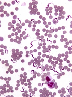
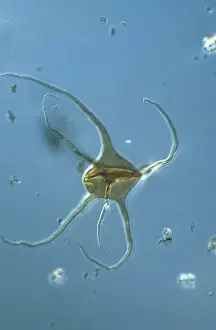
TrypanosomesScanning electron microscope image showing a trypanosoma blood smear. They have proved to be of great interest as they have evolved very differently to other better studied organisms
Ceratolithoides aculeus, coccolithScanning electron microscope image of an isolated coocolith from Cretaceous chalk. These are thin calcite shells protecting the coccolithophore within
Florosphaera profunda, coccolithScanning electron microscope image of a complete sphere of coccoliths from modern oceans. These are thin calcite shells protecting the coccolithophore within
Ophioctenella sp. brittle starScanning electron microscope image of the post-larval stage of a brittle star (x 110) A newly described species 1994
Rusty screw
Surface of a rusty screw
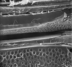
MatchstickScanning electron microscope (SEM) image showing the fractured surface of a matchstick (x 400 on a standard 9 cm wide print)
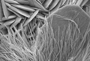
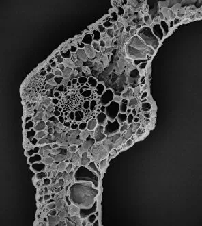
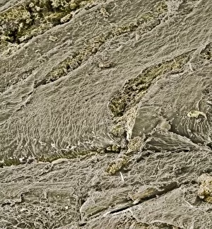
Blade of grass from a cowScanning electron microscope image of a blade of grass from a cows stomach (x 175 on a standard 9 cm wide print)
Photomicrograph: Acanthomorph acritarch, microfossil
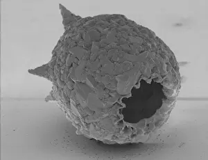
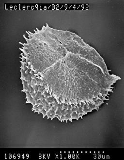
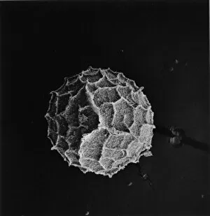
Selaginella kraussiana, spikemossScanning electron microscope image of the female spore of Krauss spikemoss (x 150 on a standard 9 cm wide print)
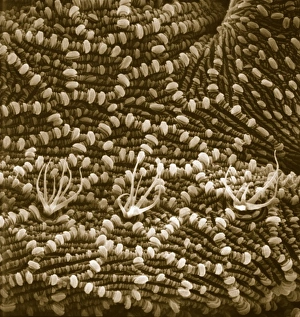
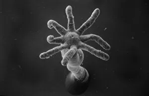
Hydra spScanning electron microscope (SEM) image showing the stinging tentacles and mouth of the coelenterate Hydra (x 36 on a standard 9cm wide print)
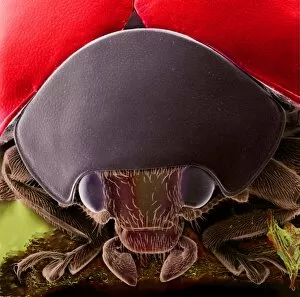
Coccinella, black spotted ladybirdScanning electron microscope image showing the head of a black spotted ladybird (x 9 on a standard 9cm wide print). Coloured artificially by computer
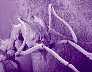
Atta cethalotes, leaf-cutter antScanning electron microscope image of a leaf-cutter ant displayed in the Darwin Centre, at the Natural History Museum, London
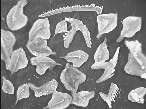
Conodont fossilsScanning electron microscope image of fossils from the Devonian period of northern Estonia, about 465 Ma old ( x 4.2). These creatures are still a mystery to paleontologists
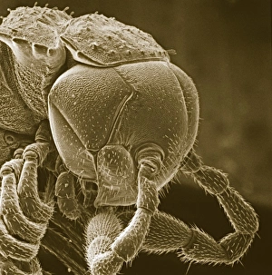
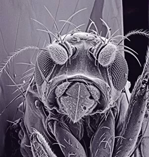
Small fly, species unknownScanning electron microscope (SEM) of a fly head. Image displayed on the glass screens in the Darwin Centre, at the Natural History Museum, London
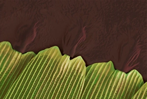
Pelargonium sp. geraniumScanning Electron Microscope image of a pelaronium leaf
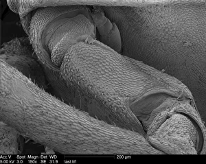
Woodlouse antennaScanning Electron Microscope (SEM) image of woodlouse antenna
OatsA scanning electron microscope (SEM) image of oats, artificially coloured by computer
Emiliania huxleyi coccosphereCoccosphere of Emiliania huxleyi from the Western Mediterranean. E. huxleyi is one of the most widespread species on earth
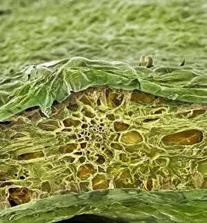
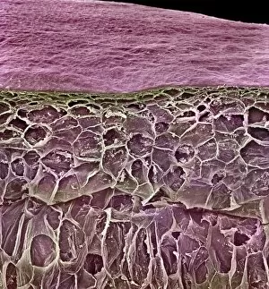
Spinacia oleracea, spinachA scanning electron microscope (SEM) image of spianch (Spinacia oleracea), artificially coloured by computer
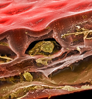
Solanum sp. tomatoA scanning electron microscope (SEM) image of a tomato (Solanum sp.), artificially coloured by computer
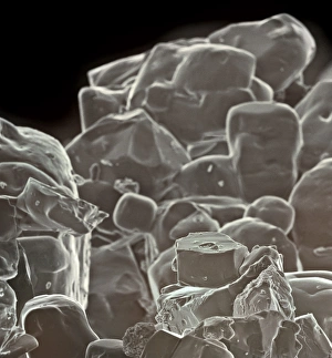
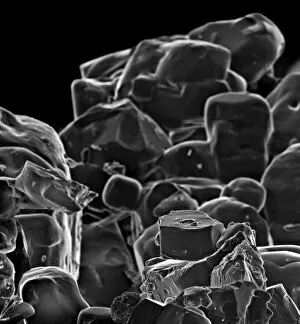
Table saltA scanning electron microscope (SEM) image of table salt, artificially coloured by computer
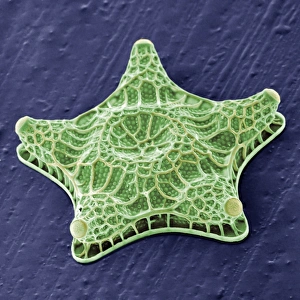
Amphitetras, diatomScanning electron microscope (SEM) image showing the diatom Amphitetras with its ornate silica shell (x5000 on a standard 9 cm wide print). Coloured artificially by computer
Vitis sp. red grapeA scanning electron microscope (SEM) image of a red grape (Vitis sp.), artificially coloured by computer
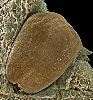
Vitis sp. grape seedScanning electron microscope (SEM) image of a grape seed (Vitis sp.), artificially coloured by computer
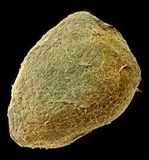
Solanum sp. tomato seedA scanning electron microscope (SEM) image of a tomato seed (Solanum sp.), artificially coloured by computer